Vasospasm vs CRAO is a triage question for every patient with sudden monocular vision loss.
Retinal artery vasospasm causes brief, repeated and reversible dimming, whereas central retinal artery occlusion (CRAO) causes abrupt, profound and persistent loss. Because CRAO behaves like an ischemic stroke, the distinction sets the pace of care.
Two Different Mechanisms
Vasospasm is a temporary constriction of the retinal or ophthalmic artery, and flow returns once the spasm relaxes, so the retina usually recovers. CRAO, however, reflects a fixed obstruction, most often an embolus or thrombus, and giant cell arteritis can also occlude the artery through vessel wall inflammation.
History Clues in Vasospasm vs CRAO
First, ask how long the episode lasted and whether it has happened before. Vasospasm typically produces stereotyped attacks lasting seconds to minutes in the same eye, and some patients link them to exercise or migraine.
By contrast, CRAO patients describe one sudden event without full recovery, and you should also ask about jaw claudication and scalp tenderness in anyone over 50.
Fundus Explorer Pro
Photograph the retinal findings described here with the phone already in your pocket — 22 D optics and built-in illumination in one handheld unit.
From Choroida — the team behind this sitePatient Profile
Vasospasm tends to affect younger adults, often with a migraine history, while CRAO usually affects older patients with hypertension, carotid disease or atrial fibrillation. However, age alone cannot settle the question, because young patients also develop embolic occlusion.
Acuity and Pupils in Vasospasm vs CRAO
Between attacks, vasospasm patients usually have normal acuity and normal pupils. In contrast, CRAO typically reduces acuity to counting fingers or worse, and a relative afferent pupillary defect also appears early and persists. However, a patent cilioretinal artery can preserve central acuity despite CRAO.
Fundus Findings in Vasospasm vs CRAO
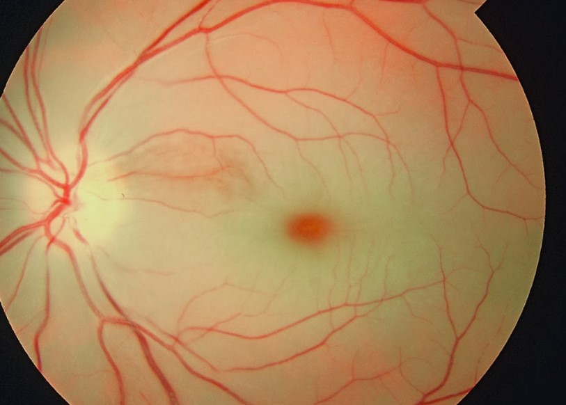
The fundus is often normal in vasospasm, since the attack has usually ended, but when an examiner catches an episode, arterioles may look narrowed, then reopen.
CRAO shows inner retinal whitening, most marked in the posterior pole. The fovea keeps its color, which creates the cherry-red spot, and you should also look for box-carring and attenuated arterioles.
Emboli as a Clue
A plaque at an arteriolar bifurcation strongly favors an embolic mechanism. Cholesterol plaques look bright and refractile, while calcific emboli look dull white. However, a missing embolus does not exclude CRAO (see retinal emboli types).
OCT in Vasospasm vs CRAO
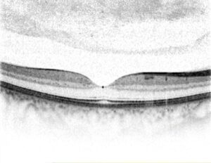
OCT gives the most objective separation in the first days, and acute CRAO shows thickened, hyperreflective inner retinal layers. Over weeks, these layers then thin and lose their normal lamination. By contrast, OCT after a brief vasospastic attack is usually normal, although prolonged spasm may rarely leave middle retinal hyperreflective bands.
Angiography Findings
Fluorescein angiography in CRAO shows delayed arterial filling and slow arteriovenous transit, and OCT angiography may also show reduced capillary flow. In vasospasm, both tests are usually normal after the episode. Therefore, a normal study supports a transient event but cannot prove its cause.
Workup in Vasospasm vs CRAO
CRAO needs the same urgent evaluation as a stroke, which means same-day referral for brain and vascular imaging and cardiac monitoring (see retinal artery occlusion workup). Patients over 50 also need ESR and CRP to screen for giant cell arteritis. Transient loss deserves the same urgency at first, because embolic amaurosis fugax looks identical.
Why Vasospasm Is a Diagnosis of Exclusion
Embolic amaurosis fugax, arteritis and hypercoagulable states can all mimic spasm. Therefore, label an episode vasospastic only after a negative workup, and recurrent identical attacks in a young migraine patient then support the diagnosis.
Treatment in Vasospasm vs CRAO
For CRAO, no ocular maneuver has proven benefit in controlled trials, and intravenous thrombolysis within a short window is still under study and belongs to stroke teams. Secondary prevention then targets the embolic source and vascular risk factors.
In vasospasm, small reports describe fewer attacks with calcium channel blockers such as nifedipine. However, that evidence is limited, so start them only after cardiovascular review.
Prognosis in Vasospasm vs CRAO
Brief vasospastic attacks usually leave no permanent damage, while CRAO, by contrast, often leaves severe permanent loss, although some eyes improve spontaneously. Patients with CRAO also carry a higher short-term risk of cerebral stroke, and for background, read idiopathic retinal vasospasm.
What to Tell the Patient
Explain that a transient attack can warn of stroke, even when vision returns fully, and then advise emergency care for any new or persisting loss. For CRAO, discuss realistic visual expectations and stroke prevention, and finally recommend general medical follow-up for both conditions.
Follow-Up
Ask vasospasm patients to keep a diary of episodes. For CRAO, check the iris and angle for neovascularization over the following months, and also repeat OCT to document inner retinal thinning.
Pitfalls to Avoid
- Do not call a first transient episode vasospasm without a full workup
- Never delay stroke referral for clinic imaging
- Do not miss giant cell arteritis in older patients
- Do not exclude CRAO because no embolus is visible


Document what you see
Two smartphone imaging tools built for everyday clinic use — one for the slit lamp, one for the fundus.
From Choroida — the team behind this siteReferences
- Hayreh SS, Zimmerman MB. Central retinal artery occlusion: visual outcome. Am J Ophthalmol. 2005;140:376-391.
- Mac Grory B, Schrag M, Biousse V, et al. Management of central retinal artery occlusion: a scientific statement from the American Heart Association. Stroke. 2021;52:e282-e294.
- Winterkorn JM, Kupersmith MJ, Wirtschafter JD, Forman S. Brief report: treatment of vasospastic amaurosis fugax with calcium-channel blockers. N Engl J Med. 1993;329:396-398.
- Varma DD, Cugati S, Lee AW, Chen CS. A review of central retinal artery occlusion: clinical presentation and management. Eye. 2013;27:688-697.
- Biousse V, Nahab F, Newman NJ. Management of acute retinal ischemia: follow the guidelines! Ophthalmology. 2018;125:1597-1607.
Test yourself
A few questions straight from this article.
-
What mechanism underlies retinal artery vasospasm, in contrast to central retinal artery occlusion?
Vasospasm is a temporary constriction of the retinal or ophthalmic artery, so flow returns when the spasm relaxes; CRAO reflects a fixed obstruction, usually an embolus or thrombus. -
Which history pattern is typical of retinal artery vasospasm rather than central retinal artery occlusion?
Vasospasm produces repeated, stereotyped attacks lasting seconds to minutes in the same eye, sometimes linked to exercise or migraine, whereas CRAO is one sudden event without full recovery. -
Which acuity and pupil findings are typical of acute central retinal artery occlusion?
CRAO typically drops acuity to counting fingers or worse and causes a relative afferent pupillary defect that appears early and persists; vasospasm patients are usually normal between attacks. -
What produces the cherry-red spot seen in central retinal artery occlusion?
CRAO whitens the inner retina, most markedly in the posterior pole, while the fovea keeps its colour, creating the cherry-red spot. -
How does a cholesterol embolus typically appear at a retinal arteriolar bifurcation?
Cholesterol plaques look bright and refractile while calcific emboli look dull white; a plaque at a bifurcation strongly favours an embolic mechanism. -
What does OCT typically show in the inner retina in acute central retinal artery occlusion?
Acute CRAO shows thickened, hyperreflective inner retinal layers on OCT, which then thin and lose their lamination over weeks. -
Which blood tests should a patient over 50 with central retinal artery occlusion have to screen for giant cell arteritis?
Alongside urgent stroke-type evaluation, patients over 50 with CRAO need ESR and CRP to screen for giant cell arteritis. -
When is it appropriate to label transient monocular vision loss as retinal vasospasm?
Vasospasm is a diagnosis of exclusion because embolic amaurosis fugax, arteritis and hypercoagulable states mimic it; recurrent identical attacks in a young migraine patient then support it. -
Which drug class has small reports of reducing attacks of retinal vasospasm?
Small reports describe fewer vasospastic attacks with calcium channel blockers such as nifedipine, but the evidence is limited, so they are started only after cardiovascular review. -
What should be checked over the months following central retinal artery occlusion?
Follow-up after CRAO includes checking the iris and angle for neovascularization and repeating OCT to document inner retinal thinning.

Writes and reviews the clinical articles on Educate. Every post is checked against peer-reviewed sources before it goes live.