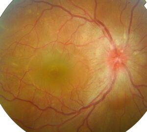
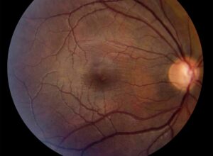
RD risk factors help clinicians decide which patients need a careful peripheral exam and clear warning advice.
Rhegmatogenous retinal detachment affects roughly 1 in 10,000 people each year, although rates vary between populations. Most cases follow a posterior vitreous detachment that tears the retina at a point of firm adhesion. Therefore, any factor that changes the vitreous or weakens the peripheral retina raises the risk.
How RD Risk Factors Act
Retinal breaks form when vitreous traction exceeds the strength of the retina, and then liquefied vitreous passes through the break and lifts the neurosensory retina. Some factors increase traction, such as early vitreous liquefaction in myopia. Others thin the peripheral retina, as lattice degeneration does, so several risks often combine in the same eye.
Myopia: The Strongest Common RD Risk Factor
Myopia accounts for a large share of detachments in phakic eyes, and in the Eye Disease Case-Control Study, risk rose with the degree of myopia. For example, eyes above 3 diopters showed roughly a tenfold higher risk than nonmyopic eyes.
Longer eyes also develop earlier vitreous liquefaction and more peripheral degeneration, while refractive surgery changes the cornea and leaves this risk unchanged. The article on retinal detachment in high myopia covers repair issues.
Fundus Explorer Pro
Photograph the retinal findings described here with the phone already in your pocket — 22 D optics and built-in illumination in one handheld unit.
From Choroida — the team behind this sitePseudophakia and Cataract Surgery
Cataract surgery raises the risk of detachment over many years after the operation, and because lens volume falls, the vitreous moves forward and may separate earlier. Risk is higher in young men, long eyes and cases with posterior capsule rupture. A whole-population study from Western Australia linked these factors to later detachment, and breaks in these eyes are often small and anterior (see pseudophakic retinal detachment).
Lattice Degeneration as an RD Risk Factor
Lattice degeneration appears in roughly 6 to 8 percent of the general population, but it occurs in a much larger share of eyes with detachment. Byer’s long-term follow-up found that few untreated lattice lesions progressed to detachment.
Therefore, lattice alone rarely justifies treatment in an asymptomatic eye. Its weight rises with myopia, a fellow-eye history or traction at the lesion edge.
Posterior Vitreous Detachment Symptoms
Acute flashes and floaters mark the period of highest short-term risk. In a JAMA meta-analysis, about one in seven patients with symptomatic PVD had a retinal tear, and vitreous hemorrhage or pigment in the anterior vitreous raised that likelihood further. As a result, these patients need a prompt dilated exam with scleral depression.
Fellow Eye Detachment
A detachment in one eye raises the risk in the other, because the fellow eye often shares the same axial length, lattice and vitreous behavior. Therefore, examine it carefully at presentation and at each follow-up visit. The post on fellow eye retinal detachment risk discusses prophylaxis decisions.
Trauma Among RD Risk Factors
Blunt trauma causes a meaningful proportion of detachments, especially in young males. Typical lesions include retinal dialysis, often inferotemporal or superonasal, and open globe injuries also carry a high risk through vitreous incarceration and proliferation. Therefore, record trauma history and examine the periphery after any significant blow.
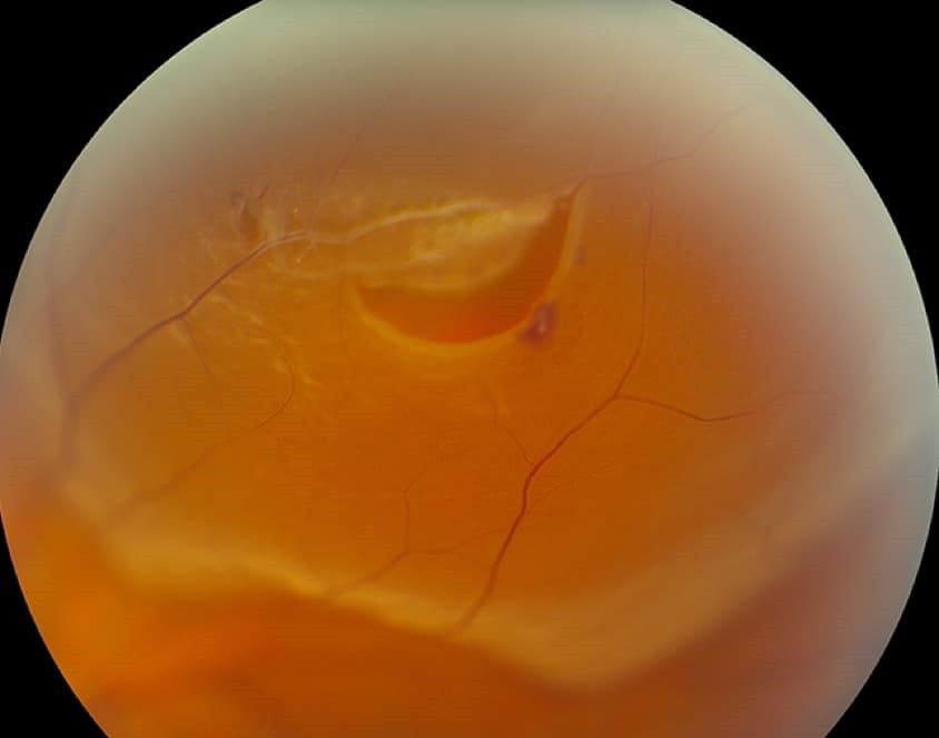
Family History and Hereditary Vitreoretinopathy
A family history of detachment increases personal risk, even without a named syndrome, but a strong pedigree should raise suspicion of Stickler syndrome. Look for high myopia, an optically empty vitreous, cleft palate, hearing loss and joint problems, and affected children need early and repeated peripheral examination.
Ranking RD Risk Factors in Practice
A practical order helps when clinic time is limited: first, acute symptoms with vitreous pigment or hemorrhage; next, a fellow-eye detachment, pseudophakia in a long eye, and high myopia. Then lattice, trauma history and family history, weighed together; finally, combinations of factors matter more than any single one.
Screening Patients With RD Risk Factors
Screening means a dilated exam of the full periphery with scleral depression. Wide-field imaging helps documentation but may miss small anterior breaks, so use it alongside the clinical exam.
Prophylactic Treatment
Laser or cryotherapy is standard for symptomatic horseshoe tears, while for asymptomatic lattice or atrophic holes, the AAO guideline generally favors observation. Treatment may still suit selected high-risk eyes, such as a fellow eye after detachment, although laser cannot prevent new breaks in untreated retina.
Counseling Patients About RD Risk Factors
Explain the warning symptoms in plain words: new floaters, flashes and a curtain or shadow, and ask patients to report them the same day. Tell myopic and pseudophakic patients that their risk persists for life.
Follow-Up After a Negative Exam
A normal first exam does not end the risk, and many clinicians repeat the exam within weeks after symptomatic PVD, especially with pigment or hemorrhage. Then routine dilated checks suit most patients with stable risk factors.
Pitfalls to Avoid
- Do not skip scleral depression in symptomatic patients
- Do not assume refractive surgery removes myopic risk
- Never treat all lattice lesions automatically


Document what you see
Two smartphone imaging tools built for everyday clinic use — one for the slit lamp, one for the fundus.
From Choroida — the team behind this siteReferences
- Eye Disease Case-Control Study Group. Risk factors for idiopathic rhegmatogenous retinal detachment. Am J Epidemiol. 1993;137:749-757.
- Hollands H, Johnson D, Brox AC, et al. Acute-onset floaters and flashes: is this patient at risk for retinal detachment? JAMA. 2009;302:2243-2249.
- Clark A, Morlet N, Ng JQ, et al. Risk for retinal detachment after phacoemulsification: a whole-population study of cataract surgery outcomes. Arch Ophthalmol. 2012;130:882-888.
- Byer NE. Long-term natural history of lattice degeneration of the retina. Ophthalmology. 1989;96:1396-1402.
- Flaxel CJ, Adelman RA, Bailey ST, et al. Posterior Vitreous Detachment, Retinal Breaks, and Lattice Degeneration Preferred Practice Pattern. Ophthalmology. 2020;127:P146-P181.

Writes and reviews the clinical articles on Educate. Every post is checked against peer-reviewed sources before it goes live.