Bacillary layer detachment is an OCT sign in which the photoreceptor layer appears split within the inner segments.
Clinicians often abbreviate it as BALAD, and because it can resemble ordinary subretinal fluid, a quick scan may miss it. Its presence also directs attention toward inflammatory and exudative disease.
What Bacillary Layer Detachment Means
The term comes from classic histology, where “bacillary layer” described the rod and cone inner and outer segments. Mehta and colleagues applied it to an OCT finding in toxoplasmosis chorioretinitis with pachychoroid disease.
Later, Cicinelli and colleagues described the sign across several posterior segment diseases. Therefore, the term names a plane of separation and does not identify one disease.
Where the Split Occurs
Ramtohul and colleagues reviewed multimodal imaging and histologic evidence, then proposed a split at the inner segment myoid, a zone that lies just beneath the external limiting membrane (ELM).
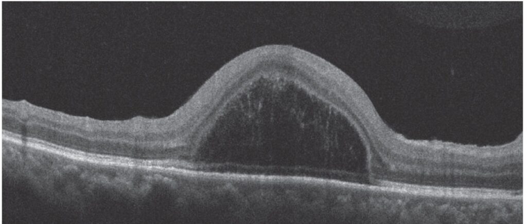
As a result, the roof carries the inner retina and ELM, while the floor keeps the ellipsoid and outer segment material on the RPE. Rapid fluid accumulation from the choroid or neovascular tissue may drive the split, but the exact sequence remains unproven.
Fundus Explorer Pro
Photograph the retinal findings described here with the phone already in your pocket — 22 D optics and built-in illumination in one handheld unit.
From Choroida — the team behind this siteOCT Features of Bacillary Layer Detachment
A well-defined hyporeflective cavity appears beneath the ELM, and its floor typically looks irregular and hyperreflective, because photoreceptor material stays on the RPE. Fine septa may also cross the cavity, particularly in acute Vogt-Koyanagi-Harada (VKH) disease.
Bacillary Layer Detachment vs Subretinal Fluid
First, trace the ELM and ellipsoid zone from healthy retina into the lesion. In ordinary fluid, the whole photoreceptor layer lifts off the RPE (see subretinal fluid on OCT).
In contrast, the BALAD cavity lies inside the photoreceptor layer, with irregular material on its floor. Both may coexist, so look for two stacked compartments.
Other OCT Lookalikes
Intraretinal cysts sit above the ELM, whereas BALAD lies below it. A pigment epithelial detachment elevates the RPE band itself. Finally, retinoschisis separates layers above the ELM and leaves the photoreceptor band more continuous.
Differential Diagnosis of Bacillary Layer Detachment
Central serous chorioretinopathy usually shows ordinary subretinal fluid, but severe cases may show a split. Therefore, correlate the cavity with angiography before naming the cause. In addition, consider choroidal tumors and other exudative lesions, because they can lift the retina.
Bacillary Layer Detachment in Inflammatory Disease
Acute VKH disease is among the best-described associations, and reports also include sympathetic ophthalmia, posterior scleritis and toxoplasmosis. Therefore, check for vitritis, disc edema, choroidal thickening and chorioretinal lesions.
Bacillary Layer Detachment in Exudative Macular Disease
Neovascular age-related macular degeneration can produce the sign, and so can polypoidal choroidal vasculopathy. For example, a new split above an irregular pigment epithelial detachment should prompt a search for active neovascularization. OCT angiography can then help confirm flow (see OCT angiography).
Clinical Presentation
Patients usually describe blurred central vision, distortion or a central scotoma. However, headache, tinnitus or neck stiffness should raise suspicion of VKH disease. On biomicroscopy, the fundus may show only a shallow serous elevation, so the diagnosis depends on OCT through the fovea.
Examination Pearls
First, ask about recent systemic symptoms, because they steer the workup. Then compare both eyes, since bilateral lesions favor inflammatory disease, and also review prior scans, because change over time clarifies the cause.
Workup After Finding Bacillary Layer Detachment
Dense OCT raster scans should cover the entire lesion, and enhanced depth imaging can then show choroidal thickness, while fluorescein and indocyanine green angiography assess leakage and polyps. En face OCT may also map the extent of the outer retinal change, and next, systemic testing should follow according to the suspected cause.
Management of Bacillary Layer Detachment
No treatment targets the sign itself, so management addresses the underlying disease. For example, VKH disease generally needs prompt systemic corticosteroids, often with immunomodulatory therapy, while neovascular disease usually receives intravitreal anti-VEGF injections.
No trial has tested treatment specifically for this sign, and risks follow the chosen therapy, including steroid complications and rare endophthalmitis. Treatment can dry the cavity, but it cannot guarantee recovery of damaged photoreceptors.
Follow-Up on OCT
Repeat scans at the same location, because small shifts can mimic change. Reports describe resolution of the cavity after effective treatment of the cause. Then assess ELM and ellipsoid zone continuity, since these bands inform visual potential.
Counseling Patients With Bacillary Layer Detachment
Explain that the scan shows a split within the light-sensing cells of the retina, and also explain that the visual outlook depends mainly on the cause and the speed of treatment.
Pitfalls to Avoid
- Do not label every hyporeflective macular space as subretinal fluid
- Avoid judging the layers from a single scan through the lesion
- Never treat the sign before identifying its likely cause
- Avoid promising visual recovery because the cavity has resolved


Document what you see
Two smartphone imaging tools built for everyday clinic use — one for the slit lamp, one for the fundus.
From Choroida — the team behind this siteReferences
- Mehta N, Chong J, Tsui E, et al. Presumed foveal bacillary layer detachment in a patient with toxoplasmosis chorioretinitis and pachychoroid disease. Retin Cases Brief Rep. 2021;15:391-398.
- Cicinelli MV, Giuffrè C, Marchese A, et al. The bacillary detachment in posterior segment ocular diseases. Ophthalmol Retina. 2020;4:454-456.
- Ramtohul P, Engelbert M, Malclès A, et al. Bacillary layer detachment: multimodal imaging and histologic evidence of a novel optical coherence tomography terminology. Retina. 2021;41:2193-2207.
- Agarwal A, Freund KB, Kumar A, et al. Bacillary layer detachment in acute Vogt-Koyanagi-Harada disease: a novel swept-source optical coherence tomography analysis. Retina. 2021;41:774-783.
Test yourself
A few questions straight from this article.
-
In classic retinal histology, what did the term "bacillary layer" describe?
The OCT term borrows from histology, where the bacillary layer meant the photoreceptor inner and outer segments, so bacillary layer detachment names a split inside the photoreceptor layer. -
At what level is the split in bacillary layer detachment proposed to occur?
Ramtohul and colleagues reviewed multimodal imaging and histology and proposed a split at the inner segment myoid, which lies just beneath the external limiting membrane. -
In bacillary layer detachment on OCT, what material forms the floor of the cavity?
The roof carries the inner retina and ELM, while the floor keeps ellipsoid and outer segment material on the RPE, which is why the floor looks irregular and hyperreflective. -
Fine septa crossing a bacillary layer detachment cavity are particularly described in which disease?
The cavity is a well-defined hyporeflective space beneath the ELM, and fine septa may cross it, particularly in acute VKH disease. -
On OCT, how does ordinary subretinal fluid differ from bacillary layer detachment?
Tracing the ELM and ellipsoid zone into the lesion shows the whole photoreceptor layer lifted in ordinary fluid, whereas the BALAD cavity lies inside the photoreceptor layer with irregular material on its floor. -
Relative to the external limiting membrane, where do intraretinal cysts lie compared with bacillary layer detachment?
Intraretinal cysts sit above the ELM, whereas a bacillary layer detachment lies below it, so the ELM is the key landmark for separating them. -
In a patient with bacillary layer detachment, which systemic symptoms should raise suspicion of VKH disease?
Patients usually report blurred central vision or distortion, but accompanying headache, tinnitus or neck stiffness should raise suspicion of Vogt-Koyanagi-Harada disease. -
A new bacillary layer detachment above an irregular pigment epithelial detachment should prompt a search for what?
Neovascular AMD and polypoidal choroidal vasculopathy can produce the sign, so a new split above an irregular PED should prompt a search for active neovascularization, with OCT angiography to help confirm flow. -
How is bacillary layer detachment managed once identified on OCT?
No treatment targets the sign itself; management addresses the cause, such as prompt systemic corticosteroids for VKH disease or intravitreal anti-VEGF for neovascular disease. -
After a bacillary layer detachment resolves, which OCT features inform visual potential?
Treatment can dry the cavity without guaranteeing photoreceptor recovery, so follow-up scans assess ELM and ellipsoid zone continuity, which inform visual potential.

Writes and reviews the clinical articles on Educate. Every post is checked against peer-reviewed sources before it goes live.