Panuveitis is intraocular inflammation involving the anterior chamber, vitreous and retina or choroid without a predominant site.
It often threatens vision because the macula and optic nerve are frequently involved. Because the differential is broad, a targeted workup saves time and avoids blind test panels.
What Panuveitis Means Under SUN
The SUN Working Group classifies uveitis by the primary site of inflammation, and panuveitis applies when the anterior chamber, vitreous and retina or choroid are all inflamed. Therefore, spillover cells during anterior uveitis do not qualify (see SUN anatomic grading), and the label describes anatomy only, so the cause still needs a search.
Symptoms Patients Report
Most patients describe blurred vision, floaters and photophobia, often in both eyes, while pain and redness vary with anterior chamber activity. In addition, headache, tinnitus or hearing changes point toward Vogt-Koyanagi-Harada (VKH) disease.
All-fit Slit-Lamp Adapter
Record and share exactly what you see at the slit lamp. One adapter fits any slit lamp or surgical microscope — and any smartphone.
From Choroida — the team behind this siteSlit Lamp and Fundus Findings in Panuveitis
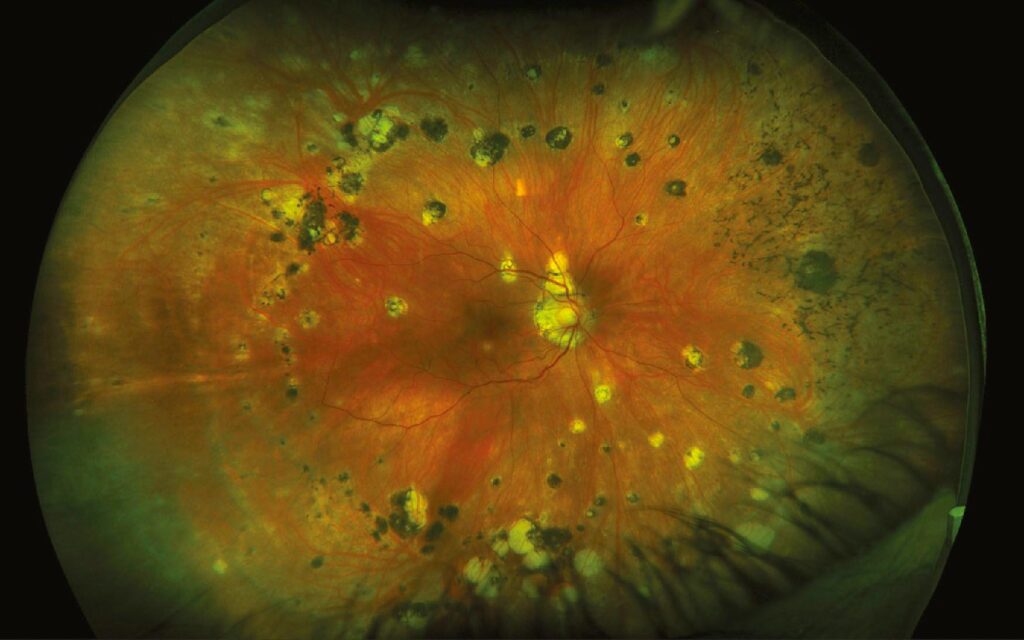
Mutton-fat keratic precipitates and iris nodules suggest a granulomatous cause. However, Behçet disease usually produces nongranulomatous inflammation, sometimes with a shifting hypopyon, so on the fundus, record vitritis, choroidal lesions, vasculitis, disc swelling and serous detachments.
Vogt-Koyanagi-Harada Disease
VKH is a bilateral granulomatous panuveitis directed against melanocytes, and the acute stage shows multiple serous retinal detachments, disc hyperemia and choroidal thickening. Meningismus and tinnitus may precede eye symptoms, while vitiligo and poliosis usually appear later. A history of penetrating trauma or intraocular surgery excludes the diagnosis.
Sympathetic Ophthalmia
Sympathetic ophthalmia follows penetrating injury or surgery to one eye, and both eyes then develop a granulomatous panuveitis that closely mimics VKH. Therefore, ask about prior trauma or vitrectomy, even years earlier (see VKH vs sympathetic ophthalmia).
Behçet Panuveitis
Behçet uveitis is recurrent and explosive, with occlusive retinal vasculitis and retinal hemorrhages, and because each attack damages the retina, delayed treatment costs vision. The diagnosis is clinical, so ask about oral and genital ulcers and skin lesions.
Sarcoidosis as a Cause of Panuveitis
Sarcoidosis produces iris nodules, vitreous snowballs, periphlebitis and multifocal choroidal lesions. Chest imaging is a high-yield screen, because hilar adenopathy supports the diagnosis, while serum ACE and lysozyme add modest support and tissue biopsy confirms it.
Tuberculosis
Tuberculous uveitis may cause serpiginous-like choroiditis, choroidal tubercles or occlusive vasculitis, and an interferon-gamma release assay plus chest imaging forms the usual screen. However, a positive assay shows exposure only, so interpret it in clinical context.
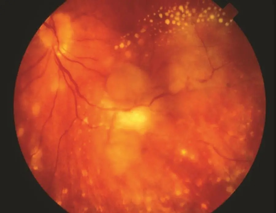
Syphilis in Panuveitis
Syphilis can mimic almost any uveitis pattern, including panuveitis, and placoid chorioretinitis and punctate inner retinitis are useful clues. Therefore, test every patient, and add HIV testing (see ocular syphilis). Treatment follows neurosyphilis regimens, so involve infectious disease colleagues.
Masquerades and Other Infections
Toxoplasmosis, viral retinitis and endogenous endophthalmitis can inflame every layer of the eye, and in older adults, vitreoretinal lymphoma may mimic chronic panuveitis with poor steroid response. When the picture does not fit, vitreous sampling becomes the next step.
History That Narrows the List
- Recent eye surgery or penetrating injury: sympathetic ophthalmia
- Headache, tinnitus, vitiligo or poliosis: VKH
- Recurrent oral and genital ulcers: Behçet disease
- Cough, dyspnea or erythema nodosum: sarcoidosis
- TB contact or endemic residence: tuberculosis
- Rash, sexual risk or HIV infection: syphilis
First-Line Panuveitis Workup
First, order syphilis serology, an interferon-gamma release assay and chest imaging for most patients, and also check a blood count, renal and liver function as a baseline before immunosuppression. Then add ACE and lysozyme when sarcoid is plausible, and finally reserve HLA typing, lumbar puncture and biopsy for specific clinical questions.
Imaging in the Workup
OCT shows macular edema and the serous detachments of acute VKH, while fluorescein angiography reveals vascular leakage, occlusion and pinpoint choroidal leaks. In addition, indocyanine green angiography detects choroidal lesions the fundus exam misses, and B-scan ultrasound helps when media opacity hides the posterior pole.
When to Start Treatment for Panuveitis
Exclude infection before high-dose corticosteroids whenever possible. Systemic steroids then control noninfectious inflammation quickly, but long-term use causes cataract, glaucoma and systemic toxicity, and therefore chronic cases usually need steroid-sparing therapy such as antimetabolites or adalimumab. In the VISUAL I trial, adalimumab lowered the risk of treatment failure in active noninfectious uveitis.
Counseling the Patient
Explain that panuveitis describes location, and the cause will guide treatment. Patients should expect several tests before a firm diagnosis, and they should also know that therapy often lasts months or years. Finally, advise urgent review for new floaters, pain or reduced vision.
Pitfalls to Avoid in Panuveitis
- Do not start high-dose steroids before syphilis and TB screening when avoidable
- Never skip asking about prior eye surgery or trauma
- Do not label disease idiopathic after an incomplete workup
- Suspect lymphoma when inflammation resists steroids in older adults


Document what you see
Two smartphone imaging tools built for everyday clinic use — one for the slit lamp, one for the fundus.
From Choroida — the team behind this siteReferences
- Jabs DA, Nussenblatt RB, Rosenbaum JT; SUN Working Group. Standardization of uveitis nomenclature for reporting clinical data. Am J Ophthalmol. 2005;140:509-516.
- Read RW, Holland GN, Rao NA, et al. Revised diagnostic criteria for Vogt-Koyanagi-Harada disease. Am J Ophthalmol. 2001;131:647-652.
- International Team for the Revision of the International Criteria for Behçet’s Disease. The International Criteria for Behçet’s Disease (ICBD). J Eur Acad Dermatol Venereol. 2014;28:338-347.
- Jaffe GJ, Dick AD, Brézin AP, et al. Adalimumab in patients with active noninfectious uveitis. N Engl J Med. 2016;375:932-943.
- Bowling B. Kanski’s Clinical Ophthalmology: A Systematic Approach. 8th ed. Elsevier. 2016.

Writes and reviews the clinical articles on Educate. Every post is checked against peer-reviewed sources before it goes live.