Blunt eye trauma compresses the globe along its front-to-back axis and stretches it at the equator.
As a result, tissues tear far from the point of impact.
Therefore, a quiet-looking eye can still hide a hyphema, a recessed angle or a retinal dialysis.
This checklist moves from the orbit to the optic nerve.
First Rule Out an Open Globe
First, decide whether the globe is intact before any pressure check.
Look for a peaked pupil, uveal prolapse, an abnormal chamber depth and 360 degree hemorrhagic chemosis.
In addition, very low pressure and restricted motility raise suspicion.
If rupture seems possible, shield the eye, avoid pressure and arrange orbital CT and surgical exploration.
History in Blunt Eye Trauma
Ask what struck the eye, how fast and from which direction.
Also ask about hammering metal on metal, because a tiny intraocular foreign body can mimic blunt injury.
Then record anticoagulant use and sickle cell status, since both change hyphema care.
All-fit Slit-Lamp Adapter
Record and share exactly what you see at the slit lamp. One adapter fits any slit lamp or surgical microscope — and any smartphone.
From Choroida — the team behind this siteVisual Acuity and Pupils
Measure acuity in each eye first, because it predicts the final outcome.
Next, test for a relative afferent pupillary defect with the swinging flashlight test.
An RAPD suggests traumatic optic neuropathy, extensive retinal detachment or dense vitreous hemorrhage.
Orbit and Lids After Blunt Eye Trauma
Palpate the orbital rims and test infraorbital sensation.
Cheek numbness with diplopia in upgaze suggests an orbital floor fracture.
In children, a white-eyed blowout with nausea and bradycardia needs urgent review, because the muscle may be entrapped.
Conjunctiva and Cornea
Isolated subconjunctival hemorrhage is usually benign.
However, bullous hemorrhagic chemosis can cover a posterior scleral rupture.
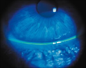
Then stain the cornea with fluorescein and check for abrasion, edema and a Seidel sign.
Anterior Chamber in Blunt Eye Trauma
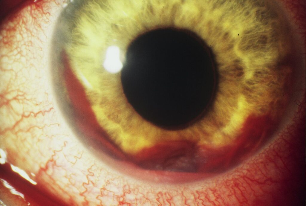
Grade cells, flare and hyphema, and compare chamber depth with the fellow eye.
Measure hyphema height in millimeters with the patient upright at every visit.
A total dark clot carries a high risk of pressure spikes and corneal blood staining (see hyphema treatment options).
Measure pressure gently and repeat it over the first days, because early spikes are common.
Iris and Pupil Signs
Sphincter tears produce small notches at the pupil margin and traumatic mydriasis.
Iridodialysis, a disinsertion of the iris root, can cause a D-shaped pupil and monocular diplopia.
Also look for iridodonesis, which hints at zonular damage.
Because sphincter damage is often permanent, counsel the patient about glare early.
Lens Assessment in Blunt Eye Trauma
Dilate once the globe is safe and the chamber allows it.
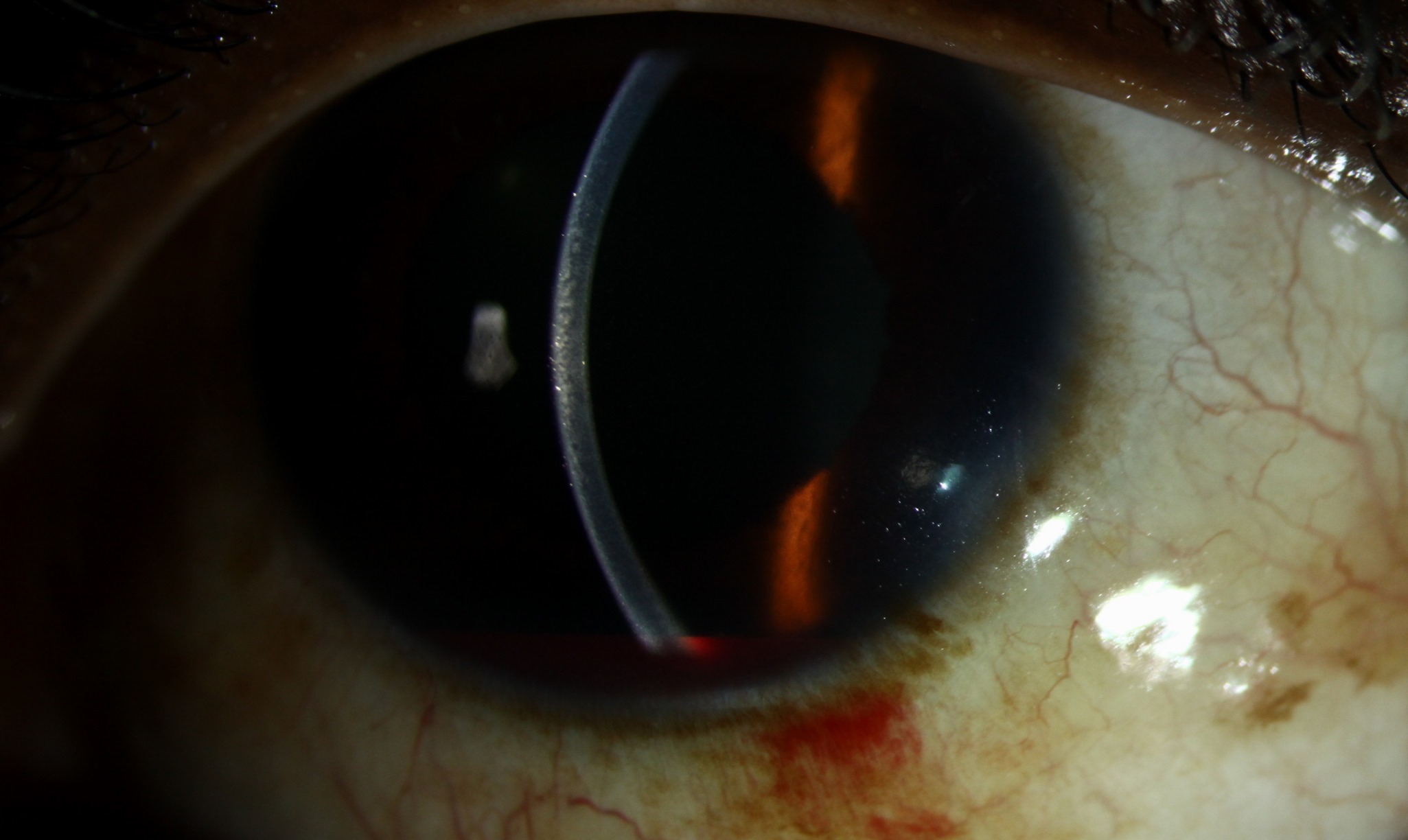
A Vossius ring of iris pigment on the anterior capsule confirms that the lens took the impact.
Then look for phacodonesis, a visible lens edge or vitreous in the chamber.
In addition, a rosette cataract may appear early or months later.
Gonioscopy and Angle Recession
Gonioscopy shows a widened ciliary body band, torn iris processes and an irregular recess.
However, delay it until any hyphema clears and rebleeding risk falls.
Angle recession raises lifelong glaucoma risk, and wider recession appears to carry more risk (see angle recession glaucoma).
As a result, these patients need long-term pressure checks even when they feel well.
Vitreous and Peripheral Retina
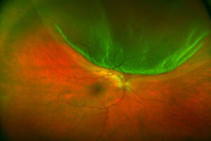
Examine the periphery with indirect ophthalmoscopy and scleral depression once you have excluded rupture.
Pigment cells in the anterior vitreous suggest a retinal break.
A superonasal retinal dialysis strongly suggests a traumatic cause.
When blood hides the fundus, B-scan ultrasound also helps exclude detachment.
Macula in Blunt Eye Trauma
Commotio retinae appears as gray-white outer retinal whitening (see commotio retinae).
OCT shows outer retinal hyperreflectivity and, in severe cases, photoreceptor disruption.
Most cases fade within weeks, while posterior pole involvement can leave pigment change.
Also check for a traumatic macular hole, which sometimes closes spontaneously in young patients.
Choroidal Rupture
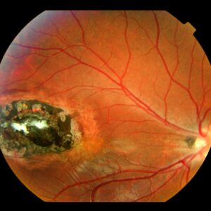
A choroidal rupture forms a yellow-white crescent concentric to the optic disc.
Overlying blood can hide it, so repeat the exam after the blood clears.
Later, choroidal neovascularization may grow from the scar, therefore teach Amsler grid self-monitoring.
Sclopetaria and the Optic Nerve
A high-velocity missile passing near the globe can tear choroid and retina without rupturing the sclera.
This chorioretinitis sclopetaria later leaves bare sclera with pigment clumping.
Next, assess disc appearance and color vision for traumatic optic neuropathy.
Evidence for high-dose corticosteroids remains weak, and the CRASH head injury trial raised safety concerns.
Follow-Up After Blunt Eye Trauma
Review hyphema patients within days for pressure and rebleeding.
Then repeat a depressed peripheral retinal exam after a few weeks.
Finally, teach every patient to report flashes, floaters or a curtain without delay.
Pitfalls to Avoid
- Never press on the globe before excluding rupture
- A floor fracture in a child with a white eye is easy to miss
- Sickle cell testing is needed in at-risk patients with hyphema
- Always plan a dilated retinal exam before discharge
- Lifelong glaucoma risk follows angle recession


Document what you see
Two smartphone imaging tools built for everyday clinic use — one for the slit lamp, one for the fundus.
From Choroida — the team behind this siteReferences
- Kuhn F, Morris R, Witherspoon CD, et al. A standardized classification of ocular trauma. Ophthalmology. 1996;103:240-243.
- Pieramici DJ, Sternberg P Jr, Aaberg TM Sr, et al. A system for classifying mechanical injuries of the eye (globe). Am J Ophthalmol. 1997;123:820-831.
- Walton W, Von Hagen S, Grigorian R, Zarbin M. Management of traumatic hyphema. Surv Ophthalmol. 2002;47:297-334.
- Sihota R, Kumar S, Gupta V, et al. Early predictors of traumatic glaucoma after closed globe injury. Arch Ophthalmol. 2008;126:921-926.

Writes and reviews the clinical articles on Educate. Every post is checked against peer-reviewed sources before it goes live.