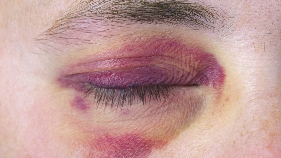
Black eye healing follows a fairly predictable sequence of color changes over two to three weeks.
The clinical term is periorbital ecchymosis, meaning blood within the loose eyelid and orbital tissues. However, the bruise tells you little about the eye beneath it. Therefore, every black eye deserves a careful look at the globe.
Why the Eyelid Bruises So Easily
Eyelid skin is the thinnest skin on the body, and the subcutaneous tissue is loose and pools blood easily. As a result, a modest blow can produce dramatic swelling. Blood also tracks with gravity, so a forehead injury may bruise the lids a day later.
Day 0 to 2 in Black Eye Healing
Fresh blood gives the lid a red or reddish purple color, and swelling peaks within the first 24 to 48 hours, when the lids may close. Then deoxygenated hemoglobin shifts the color toward deep blue or purple. Patients often worry most now, although resorption has already started.
All-fit Slit-Lamp Adapter
Record and share exactly what you see at the slit lamp. One adapter fits any slit lamp or surgical microscope — and any smartphone.
From Choroida — the team behind this siteDays 3 to 6: Blue Turns Green
Macrophages break hemoglobin down into biliverdin, a green pigment. Therefore, the edges often turn green while the center stays purple. Swelling usually settles enough for the patient to open the eye.
Days 7 to 10: The Yellow Phase
Next, biliverdin converts to bilirubin, which looks yellow. Hemosiderin then adds a brownish tint that clears slowly. However, the yellow rim may spread wider than the original bruise.
Weeks 2 to 3 in Black Eye Healing
Most bruises fade completely within two to three weeks, although older patients and those taking anticoagulants often take longer. Brown staining beyond a month is uncommon, so reconsider the diagnosis then.
Can You Date a Bruise by Color?
Color offers only a rough guide to age, and Langlois and Gresham found that yellow appeared only in bruises older than about 18 hours. However, a systematic review in children concluded that color cannot age a bruise accurately. Therefore, avoid precise dating in medicolegal or safeguarding notes.
What Speeds Black Eye Healing
First, apply cold compresses in short sessions during the first 24 to 48 hours, while head elevation during sleep also reduces dependent swelling. For pain, acetaminophen is a reasonable choice because it spares platelet function. After swelling settles, gentle warm compresses may help, although evidence is limited.
What Slows Black Eye Healing
Aspirin, anticoagulants and some supplements prolong bleeding into the tissues, and advanced age, liver disease and bleeding disorders delay clearance. Rubbing or a second injury also restarts the cycle, but patients must not stop prescribed anticoagulants without medical advice.
Myths About Black Eye Healing
Raw steak offers no benefit and carries an infection risk. Arnica and vitamin K creams are popular, while supporting evidence is limited and inconsistent, so describe them as optional and unproven.
Examining the Globe Behind a Black Eye
Open swollen lids gently, using retractors and avoiding pressure on the globe. First, check visual acuity and test pupils for a relative afferent defect. Next, examine the anterior segment at the slit lamp (see slit lamp technique), and finally measure pressure and dilate the fundus once you exclude an open globe.
Anterior Segment Signs to Look For
Hyphema or microhyphema signals intraocular bleeding and needs pressure monitoring. An irregular, poorly reactive pupil suggests traumatic mydriasis or sphincter tears. Fluorescein may also reveal a corneal abrasion under the swollen lid, and a subconjunctival hemorrhage without a visible posterior limit can hide a scleral rupture.
Orbital Fracture Clues
Diplopia on upgaze suggests inferior rectus entrapment in an orbital floor fracture, and cheek or upper lip numbness also points to infraorbital nerve injury. Crepitus after nose blowing means air has entered from a sinus. Then enophthalmos may appear only after the swelling subsides.
Warning Signs in Black Eye Healing
- Reduced vision or a dark shadow
- Double vision or painful eye movements
- A tense, proptotic orbit with rising pain
- New flashes or floaters
- Bruising around both eyes after head trauma
- A bruise that keeps enlarging, or bruising without injury
Retrobulbar Hemorrhage
A tense orbit with vision loss and high pressure suggests orbital compartment syndrome, which needs lateral canthotomy and cantholysis without delay. Therefore, do not wait for imaging when the clinical signs are clear.
Counseling During Black Eye Healing
Tell patients the colors will change, and green or yellow means resorption. Advise a prompt return for any vision change, diplopia or worsening pain. Patients with hyphema also need rest and an early pressure check, and because angle recession can raise pressure years later, arrange long-term follow-up (see same-day eye emergencies).
Pitfalls to Avoid
- Do not dismiss a black eye without checking vision and pupils
- Never force swollen lids open over a possible ruptured globe
- Avoid dating a bruise precisely by color
- Do not overlook bilateral bruising after head injury


Document what you see
Two smartphone imaging tools built for everyday clinic use — one for the slit lamp, one for the fundus.
From Choroida — the team behind this siteReferences
- Langlois NE, Gresham GA. The ageing of bruises: a review and study of the colour changes with time. Forensic Sci Int. 1991;50:227-238.
- Maguire S, Mann MK, Sibert J, Kemp A. Can you age bruises accurately in children? A systematic review. Arch Dis Child. 2005;90:187-189.
- Kuhn F, Morris R, Witherspoon CD, et al. A standardized classification of ocular trauma. Ophthalmology. 1996;103:240-243.
- American Academy of Ophthalmology. Basic and Clinical Science Course, Section 7: Oculofacial Plastic and Orbital Surgery. San Francisco: AAO; 2023.
- Salmon JF. Kanski’s Clinical Ophthalmology: A Systematic Approach. 9th ed. Elsevier; 2020.

Writes and reviews the clinical articles on Educate. Every post is checked against peer-reviewed sources before it goes live.