Iris nodules are focal elevations of the iris surface, and their causes range from hamartoma to inflammation and tumor.
Location, color, laterality and age usually narrow the differential before any test.
Therefore, a structured slit lamp description is the most useful first step.
What Iris Nodules Are
A nodule is a discrete raised lesion that interrupts the normal stromal pattern.
It may arise from melanocytes, inflammatory cells, histiocytes or tumor cells.
In addition, some lesions only look nodular, such as cysts pushing the stroma forward.
First Questions for Iris Nodules
First, start with age, because children and older adults have different typical causes.
Next, ask about pain, redness, photophobia and previous uveitis.
Then record skin findings, known cancer and family history of neurofibromatosis.
Bilateral nodules usually point to systemic disease.
All-fit Slit-Lamp Adapter
Record and share exactly what you see at the slit lamp. One adapter fits any slit lamp or surgical microscope — and any smartphone.
From Choroida — the team behind this siteLisch Nodules in NF1
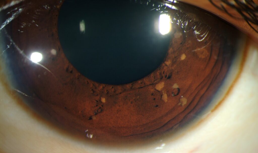
Lisch nodules are smooth, dome-shaped, tan to brown melanocytic hamartomas on the anterior iris surface.
They are typically bilateral and become more frequent with age in neurofibromatosis type 1.
Also, two or more Lisch nodules count as a diagnostic criterion in the revised NF1 criteria.
However, they do not affect vision, so they need no treatment.
Koeppe and Busacca Nodules
Koeppe nodules sit at the pupillary margin and look small, gray-white and translucent.
Busacca nodules, in contrast, lie on the iris stroma, away from the pupil.
Both suggest granulomatous uveitis, for example sarcoidosis, tuberculosis or Vogt-Koyanagi-Harada disease.
For a detailed review, see Koeppe and Busacca nodules.
Granulomas and Berlin Nodules
True iris granulomas are larger, fleshy and often vascularized.
Berlin nodules occur in the angle, so only gonioscopy reveals them.
Also, mutton-fat keratic precipitates and anterior chamber cells usually accompany these lesions.
Therefore, an inflamed eye with a nodule needs a uveitis workup first.
Juvenile Xanthogranuloma
JXG typically presents in young children as a yellow-orange or tan vascular iris lesion.
Spontaneous hyphema, heterochromia and secondary glaucoma are common presenting signs.
In addition, coexisting skin papules support the diagnosis (see juvenile xanthogranuloma).
Melanocytic Tumors as Iris Nodules
An iris nevus is a flat or mildly elevated pigmented lesion with stable borders.
Melanoma, however, tends to be thicker and more vascular, often in the inferior iris.
The ABCDEF guide flags young age, hyphema, inferior location, diffuse growth, ectropion uveae and feathery margins.
As a result, documented growth needs prompt evaluation (see iris melanoma).
Other Tumor Nodules
Iris metastases usually appear as white, pink or yellow fluffy masses that grow quickly.
Also, leukemia and lymphoma can infiltrate the iris and cause pseudohypopyon.
In children, retinoblastoma may seed the anterior chamber and mimic inflammation.
Comparing Iris Nodules by Feature
- First, Lisch: anterior surface, tan to brown, bilateral, NF1
- Next, Koeppe: pupillary margin, gray-white, granulomatous uveitis
- Then Busacca: midstroma, white to pink, granulomatous uveitis
- Also granuloma: any site, fleshy and vascular, sarcoidosis or tuberculosis
- In addition, JXG: any site, yellow-orange, young children
- However, nevus or melanoma: often inferior, brown or amelanotic, unilateral
- Finally, metastasis: any site, white to pink, known or occult carcinoma
Mimics of Iris Nodules
Pigment epithelial cysts can bulge the stroma and simulate a solid mass.
Freckles, however, are flat and do not distort the stroma.
Mammillations, in contrast, are tiny, uniform, villiform projections, often with ocular melanocytosis.
Gonioscopy and Imaging
Gonioscopy shows angle nodules, angle seeding and ciliary body extension.
Then ultrasound biomicroscopy measures thickness and separates cysts from solid lesions.
Also, baseline slit lamp photographs allow objective comparison later.
Systemic Workup for Iris Nodules
Inflammatory nodules call for chest imaging, serum ACE or lysozyme, and tuberculosis testing.
In addition, syphilis serology is reasonable in granulomatous uveitis.
For Lisch nodules, examine the skin for café-au-lait macules and arrange NF1 evaluation.
Follow-Up of Iris Nodules
Inflammatory nodules should shrink as the uveitis responds to treatment.
However, persistent nodules despite control suggest a wrong diagnosis or masquerade syndrome.
Then stable pigmented lesions need periodic photographs, with intervals based on risk.
Also check intraocular pressure at every visit, because several of these lesions raise it.
Counseling Patients
First, explain that most iris nodules are benign or inflammatory.
For uveitis, stress adherence and prompt return if pain or blurring worsens.
For suspicious pigmented lesions, explain why serial photographs matter.
When to Refer
Refer to an ocular oncologist when a lesion grows, bleeds, distorts the pupil or seeds the angle.
Also refer any child with hyphema and an iris mass.
However, biopsy is reserved for uncertain cases.
Pitfalls to Avoid
- Do not label every pigmented bump a nevus without gonioscopy
- Never treat a child’s nodules as uveitis before excluding retinoblastoma
- Also use oblique light, because Lisch nodules hide on dark irides
- Avoid calling a cyst a solid tumor without ultrasound biomicroscopy


Document what you see
Two smartphone imaging tools built for everyday clinic use — one for the slit lamp, one for the fundus.
From Choroida — the team behind this siteReferences
- Lewis RA, Riccardi VM. Von Recklinghausen neurofibromatosis: incidence of iris hamartomata. Ophthalmology. 1981;88:348-354.
- Legius E, Messiaen L, Wolkenstein P, et al. Revised diagnostic criteria for neurofibromatosis type 1 and Legius syndrome: an international consensus recommendation. Genet Med. 2021;23:1506-1513.
- Jabs DA, Nussenblatt RB, Rosenbaum JT. Standardization of uveitis nomenclature for reporting clinical data. Am J Ophthalmol. 2005;140:509-516.
- Zimmerman LE. Ocular lesions of juvenile xanthogranuloma. Trans Am Acad Ophthalmol Otolaryngol. 1965;69:412-442.
- Shields CL, Kaliki S, Hutchinson A, et al. Iris nevus growth into melanoma: analysis of 1611 consecutive eyes: the ABCDEF guide. Ophthalmology. 2013;120:766-772.

Writes and reviews the clinical articles on Educate. Every post is checked against peer-reviewed sources before it goes live.