Aponeurotic ptosis is the most common form of acquired upper eyelid drooping in adults.
It results from stretching or disinsertion of the levator aponeurosis from the tarsus, and because the muscle still works, levator function remains good. As a result, tendon repair gives predictable results in most patients.
Anatomy of Aponeurotic Ptosis
Below Whitnall’s ligament, the levator becomes a broad aponeurosis inserting on the anterior tarsus, and fine fibers also pass to the skin and form the lid crease. When the aponeurosis slips upward, the crease therefore rises with it, and the superior sulcus deepens as orbital fat retreats.
Common Causes
Aging is the leading cause, because connective tissue weakens over decades. However, younger adults develop the same picture after repeated mechanical stress.
- Involutional change with age
- Long-term rigid contact lens wear
- Intraocular or eyelid surgery
- Chronic eye rubbing
- Recurrent lid edema or blunt trauma
All-fit Slit-Lamp Adapter
Record and share exactly what you see at the slit lamp. One adapter fits any slit lamp or surgical microscope — and any smartphone.
From Choroida — the team behind this siteContact Lenses and Aponeurotic Ptosis
Rigid contact lens wear is a recognized cause in young and middle-aged adults. For example, pulling the lid sideways during lens removal may stretch the aponeurosis over years, while soft lenses appear to carry a lower risk. Therefore, ask about lens type, wear time and overwear habits (see contact lens overwear syndrome).
Ptosis After Eye Surgery
Drooping can follow cataract, glaucoma, corneal or retinal surgery, and suspected mechanisms include speculum stretch, bridle sutures and local anesthetic injection. However, mild cases often improve as swelling settles, and therefore most surgeons wait several months before repair.
What Patients Report
Patients first notice a tired appearance, and then they describe heaviness and brow ache that worsen later in the day. A marked droop also blocks the superior visual field, especially while reading, and many patients raise the brow constantly, which causes forehead fatigue.
Exam Signs of Aponeurotic Ptosis
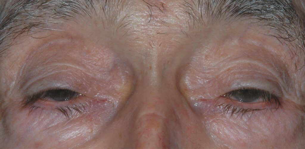
The classic picture is a low lid with a high crease.
- Good levator function, often near normal
- Elevated, faint or absent lid crease
- Thin lid tissue above the tarsus
- Deep superior sulcus
- Lid stays low in downgaze
- Raised brow because of frontalis overaction
Measuring the Lid
First, measure the margin reflex distance 1 with the brow relaxed, and next block the brow and record levator excursion from downgaze to upgaze. Also note crease height, Bell’s phenomenon and corneal staining, and finally lift the droopier lid and watch the fellow lid, because Hering’s law may unmask bilateral disease.
Differential Diagnosis
Good levator function narrows the differential but does not close it.
- Myasthenia gravis: variable droop, fatigue, diplopia
- Horner syndrome: mild ptosis with miosis
- Third nerve palsy: poor levator function and motility deficit
- Brow ptosis or dermatochalasis: a false droop (see pseudoptosis)
Any acute ptosis with pupil change or diplopia needs urgent assessment.
Phenylephrine Test in Aponeurotic Ptosis
Phenylephrine 2.5% in the upper fornix stimulates Müller’s muscle, and then the examiner remeasures the lid after several minutes. A clear lift supports a posterior approach and also reveals any Hering-related drop in the fellow lid.
Levator Advancement for Aponeurotic Ptosis
External levator advancement is the standard repair, and first the surgeon opens the crease, finds the dehisced aponeurosis and resutures it to the tarsus. Then, under local anesthesia, the patient opens the eye, so height and contour can be adjusted, and finally excess skin can be trimmed at the same time.
Müller Muscle Resection
Müller muscle conjunctival resection works from the back of the lid and suits mild ptosis with a positive phenylephrine response. In addition, it avoids a skin incision and usually gives a smooth contour.
Risks of Ptosis Surgery
Undercorrection and overcorrection are the commonest problems, and contour peaks, crease asymmetry and lagophthalmos also occur. Exposure keratopathy is a concern when Bell’s phenomenon or the tear film is poor, while levator surgery does not lift a descended brow.
Counseling in Aponeurotic Ptosis
Explain that the muscle is healthy and the tendon has loosened. Patients can expect a better field and appearance, although perfect symmetry is not guaranteed. In addition, contact lens wearers can usually continue, using gentle removal technique, so also discuss the chance of revision openly.
Follow-Up After Repair
Review the lid at one week, then after swelling settles, and at each visit also check height, contour, closure and corneal staining. Because early asymmetry often improves, delay revision until the lid stabilizes.
Pitfalls to Avoid
- Do not miss variable ptosis, because it suggests myasthenia
- Never skip the pupil and motility exam
- Test the fellow lid before surgery, since Hering’s law can hide drooping
- Avoid overcorrection when Bell’s phenomenon is poor


Document what you see
Two smartphone imaging tools built for everyday clinic use — one for the slit lamp, one for the fundus.
From Choroida — the team behind this siteReferences
- Kersten RC, de Conciliis C, Kulwin DR. Acquired ptosis in the young and middle-aged adult population. Ophthalmology. 1995;102:924-928.
- van den Bosch WA, Lemij HG. Blepharoptosis induced by prolonged hard contact lens wear. Ophthalmology. 1992;99:1759-1765.
- Jones LT, Quickert MH, Wobig JL. The cure of ptosis by aponeurotic repair. Arch Ophthalmol. 1975;93:629-634.
- Putterman AM, Urist MJ. Müller muscle-conjunctiva resection: technique for treatment of blepharoptosis. Arch Ophthalmol. 1975;93:619-623.
- Finsterer J. Ptosis: causes, presentation, and management. Aesthetic Plast Surg. 2003;27:193-204.
- American Academy of Ophthalmology. Basic and Clinical Science Course, Section 7: Oculofacial Plastic and Orbital Surgery. San Francisco: AAO; 2023.

Writes and reviews the clinical articles on Educate. Every post is checked against peer-reviewed sources before it goes live.