BRVO vs BRAO is a frequent question when a patient reports sudden sectoral vision loss in one eye.
Both affect a single retinal branch, so the field defects can look alike.
However, the fundus, OCT and urgency of the workup differ sharply.
BRVO vs BRAO: The Core Difference
A branch retinal vein occlusion blocks venous outflow from one retinal sector.
Blood and fluid then back up into the tissue upstream.
In contrast, a branch retinal artery occlusion stops arterial inflow.
As a result, the inner retina in that territory becomes ischemic.
Where Each Occlusion Starts
BRVO usually begins at an arteriovenous crossing, where a stiff artery compresses the vein.
The superotemporal quadrant is the most common site.
BRAO, however, usually follows an embolus lodged at an arterial bifurcation.
Fundus Explorer Pro
Photograph the retinal findings described here with the phone already in your pocket — 22 D optics and built-in illumination in one handheld unit.
From Choroida — the team behind this siteSymptoms in BRVO vs BRAO
Patients with BRVO often notice blurring only when macular edema reaches the fovea.
Some have no symptoms at all when the macula is spared.
By contrast, BRAO causes abrupt, painless loss of a defined part of the field.
Also, some patients report earlier transient episodes of monocular vision loss.
Fundus Clues in BRVO
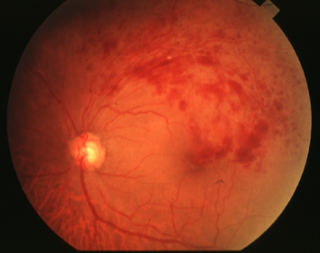
Flame and dot-blot hemorrhages fan out from the crossing into the drained sector.
In addition, dilated veins, cotton wool spots and hard exudates may appear.
Changes stay within the territory of the occluded vein (see branch retinal vein occlusion).
Fundus Clues in BRAO
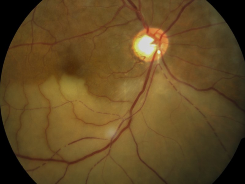
In the ischemic sector, the retina looks pale and whitened from inner retinal edema.
Sometimes the arteriole appears narrowed, with segmentation of the blood column.
Next, search the arterial tree for a refractile Hollenhorst plaque or a dull white embolus.
Hemorrhages are scarce, unlike the picture in BRVO (see branch retinal artery occlusion).
BRVO vs BRAO: Side-by-Side Signs
- Hemorrhages: abundant in BRVO, scarce in BRAO
- Retinal color: congested in BRVO, pale white in BRAO
- Vessels: dilated veins in BRVO, attenuated arterioles in BRAO
- Emboli: absent in BRVO, often visible in BRAO
- Main macular threat: edema in BRVO, inner retinal ischemia in BRAO
OCT in BRVO vs BRAO
In BRVO, OCT usually shows cystoid macular edema, often with subretinal fluid.
In acute BRAO, however, the inner retinal layers look thickened and hyperreflective.
Later, these layers thin, while the outer retina stays relatively intact.
Therefore, sectoral inner thinning can reveal an old, missed BRAO.
Also, fluorescein angiography shows delayed branch filling in BRAO and nonperfusion with leakage in BRVO.
Urgency and Triage
BRAO is an acute retinal ischemic event, similar to a transient ischemic attack or stroke.
Therefore, the patient needs same-day stroke evaluation, ideally at a stroke center.
BRVO is rarely an emergency.
However, it still needs prompt assessment for macular edema and ischemia.
Systemic Workup for BRAO
The workup searches for an embolic source.
It usually includes carotid imaging, echocardiography, rhythm monitoring and brain imaging.
In older patients, also consider giant cell arteritis when symptoms suggest it.
Follow the steps in retinal artery occlusion workup.
Systemic Workup for BRVO
Hypertension is the strongest association, followed by diabetes and hyperlipidemia.
Also, check intraocular pressure, because glaucoma raises the risk of vein occlusion.
Routine thrombophilia testing adds little in older patients with typical risk factors.
Treatment in BRVO vs BRAO
Anti-VEGF injections improved vision in BRVO macular edema in the BRAVO trial.
Grid laser, supported by the Branch Vein Occlusion Study, still helps selected persistent edema.
Then scatter laser treats neovascularization once it develops.
By contrast, no treatment has proven benefit for restoring perfusion in BRAO.
Prognosis in BRVO vs BRAO
Many BRVO eyes improve over months, as collaterals form and edema responds to treatment.
In BRAO, acuity often stays good when the fovea is spared.
However, the sectoral field defect usually persists.
Counseling the Patient
Use plain words: a blocked vein causes bleeding and swelling, while a blocked artery starves the retina.
For BRAO, explain that the eye may signal vascular risk elsewhere in the body.
For BRVO, set expectations about repeated injections and regular visits.
Follow-Up in BRVO vs BRAO
After BRVO, track edema on OCT and watch for neovascularization and vitreous hemorrhage.
After BRAO, examine the fellow eye and look for recurrent emboli.
Finally, coordinate with the primary physician to control vascular risk factors.
Pitfalls to Avoid
- Do not call a pale sector BRVO because a few hemorrhages are present
- Never delay stroke referral because visual acuity is good
- Check for cilioretinal artery involvement when whitening sits near the disc
- Remember that macular BRVO can show edema with few hemorrhages


Document what you see
Two smartphone imaging tools built for everyday clinic use — one for the slit lamp, one for the fundus.
From Choroida — the team behind this siteReferences
- Hayreh SS, Zimmerman MB. Fundus changes in branch retinal vein occlusion. Retina. 2015;35:1016-1027.
- Branch Vein Occlusion Study Group. Argon laser photocoagulation for macular edema in branch vein occlusion. Am J Ophthalmol. 1984;98:271-282.
- Campochiaro PA, Heier JS, Feiner L, et al. Ranibizumab for macular edema following branch retinal vein occlusion: six-month primary end point results of a phase III study. Ophthalmology. 2010;117:1102-1112.
- Mason JO 3rd, Shah AA, Vail RS, et al. Branch retinal artery occlusion: visual prognosis. Am J Ophthalmol. 2008;146:455-457.
- Biousse V, Nahab F, Newman NJ. Management of acute retinal ischemia: follow the guidelines! Ophthalmology. 2018;125:1597-1607.
- Flaxel CJ, Adelman RA, Bailey ST, et al. Retinal Vein Occlusions Preferred Practice Pattern. Ophthalmology. 2020;127:P288-P320.

Writes and reviews the clinical articles on Educate. Every post is checked against peer-reviewed sources before it goes live.