Trantas dots are small, chalky white spots at the limbus that signal active vernal keratoconjunctivitis (VKC).
Clinicians also call them Horner-Trantas dots, after the two physicians who described them.
They sit on a gelatinous, thickened limbus and change quickly with disease activity.
Therefore, a careful limbal examination tells you how active the allergy is today.
What Trantas Dots Are
Each dot is a tiny cluster of degenerated eosinophils and shed epithelial cells.
The cells gather at the tips of limbal papillae, where inflammation is most intense.
As a result, the dot looks raised, white and slightly gritty.
Because eosinophils drive them, the dots point to allergy, and infection does not produce them.
Who Develops Them
Trantas dots appear mainly in children and adolescents with VKC, and boys outnumber girls.
Limbal VKC also seems more frequent in warm, dry climates and in patients of African or Asian descent (see limbal vernal keratoconjunctivitis).
In addition, a personal or family history of atopy is common.
Adults with atopic keratoconjunctivitis may occasionally show similar dots.
All-fit Slit-Lamp Adapter
Record and share exactly what you see at the slit lamp. One adapter fits any slit lamp or surgical microscope — and any smartphone.
From Choroida — the team behind this siteHow Trantas Dots Look at the Slit Lamp
Use diffuse light first, then a narrow beam across the limbus.
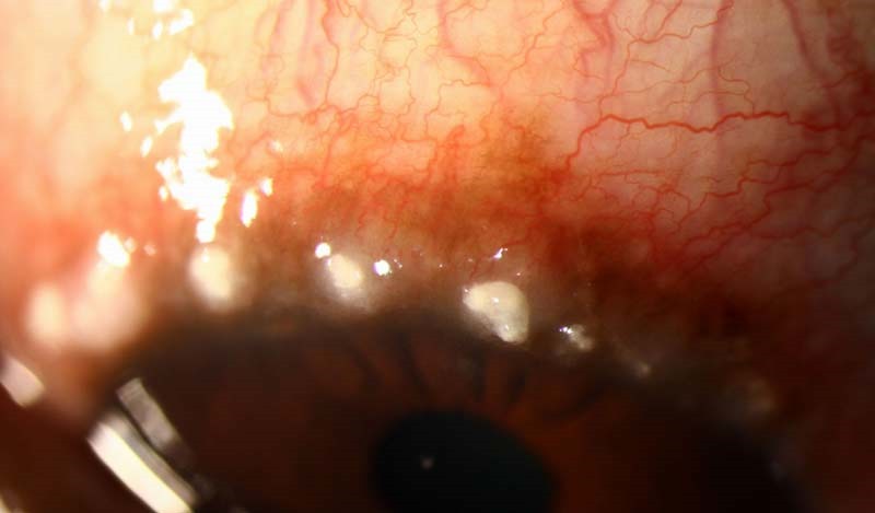
The dots appear as discrete white or yellow-white points, often in a row along the upper limbus.
They rest on a swollen, gelatinous ring that may look gray or pink.
In darker-skinned patients, brown limbal pigmentation may also surround the swelling.
Trantas Dots and VKC Activity
Trantas dots behave as a marker of active inflammation.
They tend to appear during flares and fade within days to weeks once treatment works.
Therefore, new dots support stepping up treatment, while their disappearance suggests control.
Published VKC grading systems also count limbal infiltrates and dots among activity signs.
Why They Come and Go
The dots consist of cells, with no fibrous or degenerative tissue.
As a result, they clear as eosinophils break down and the epithelium sheds.
However, persistent limbal inflammation can injure limbal stem cells over time.
Chronic, poorly controlled limbal VKC may then cause conjunctivalization of the peripheral cornea.
Associated Corneal Signs
Active limbal disease often comes with superior punctate epitheliopathy.
In addition, eosinophil products can cause macroerosions and a shield ulcer.
Pseudogerontoxon, an arcus-like scar, may follow repeated limbal flares.
Fluorescein staining helps you grade each finding at every visit.
Diagnosing Trantas Dots
The diagnosis is clinical and rests on the slit lamp picture in an allergic patient.
A conjunctival scraping, when performed, may show eosinophils and support the allergic cause.
Also, tear IgE and skin prick tests can add information about atopy.
However, no test replaces a careful limbal examination.
Mimics of Trantas Dots
Several white limbal lesions can confuse the examiner.
- Phlyctenules: raised nodules with a vascular leash, linked to staphylococcal or tuberculous hypersensitivity
- White limbal girdle of Vogt: a chalky crescent at 3 and 9 o’clock in older adults (see Vogt limbal girdle)
- Pinguecula: a yellowish interpalpebral deposit that stays stable for months
- Early pterygium, which grows onto the cornea as a vascular wing (see pterygium)
Trantas Dots Versus Phlyctenules
Phlyctenules usually appear as one or a few pinkish-white nodules with feeder vessels.
They often ulcerate at the apex and may migrate onto the cornea.
Trantas dots, by contrast, are multiple, small and avascular.
Also, phlyctenules accompany blepharitis, while Trantas dots accompany itching and tarsal papillae.
Treatment When Trantas Dots Are Present
Treatment targets VKC activity, because the dots simply follow the disease.
First, start mast cell stabilizer or dual-action antihistamine drops with cold compresses.
Next, add a short pulse of topical corticosteroid for moderate or severe flares.
Then use topical cyclosporine or tacrolimus as steroid-sparing therapy.
The VEKTIS trial supported cyclosporine cationic emulsion in children with severe VKC.
Limits and Risks of Therapy
Steroids clear the dots quickly; however, they raise intraocular pressure and promote cataract.
Therefore, check pressure at every visit during a steroid course.
Calcineurin inhibitors may sting, and they act more slowly than steroids in an acute flare.
Finally, no drug cures VKC, although many cases remit after puberty.
Counseling the Family
Explain that the white spots reflect active allergy and usually fade with treatment.
Also, stress that eye rubbing worsens inflammation and may promote keratoconus.
Warn against restarting steroid drops without supervision.
Finally, plan a review before the expected seasonal flare.
Follow-Up Signs to Record
- Number of Trantas dots and their clock hours
- Limbal swelling and pigmentation
- Superior corneal staining or a shield ulcer
- Intraocular pressure during steroid courses
- Early signs of limbal stem cell deficiency
Pitfalls to Avoid
- Never call a single vascularized nodule a Trantas dot
- Always evert the upper lid, because tarsal papillae often coexist
- Treat the corneal epithelium as well as the limbus
- Check intraocular pressure in every child on steroids


Document what you see
Two smartphone imaging tools built for everyday clinic use — one for the slit lamp, one for the fundus.
From Choroida — the team behind this siteReferences
- Bonini S, Bonini S, Lambiase A, et al. Vernal keratoconjunctivitis revisited: a case series of 195 patients with long-term followup. Ophthalmology. 2000;107:1157-1163.
- Leonardi A, Doan S, Amrane M, et al. A randomized, controlled trial of cyclosporine A cationic emulsion in pediatric vernal keratoconjunctivitis: the VEKTIS study. Ophthalmology. 2019;126:671-681.
- Kumar S. Vernal keratoconjunctivitis: a major review. Acta Ophthalmol. 2009;87:133-147.
- Singhal D, Sahay P, Maharana PK, et al. Vernal keratoconjunctivitis. Surv Ophthalmol. 2019;64:289-311.
- Bowling B. Kanski’s Clinical Ophthalmology: A Systematic Approach. 8th ed. Elsevier; 2016.

Writes and reviews the clinical articles on Educate. Every post is checked against peer-reviewed sources before it goes live.