Hyphema rebleed is new anterior chamber bleeding after a traumatic hyphema, usually within the first five days.
It often raises pressure more than the first bleed and can carry a worse visual outlook.
Therefore, the first week needs a clear monitoring plan.
What Counts as a Hyphema Rebleed
Secondary hemorrhage means new bleeding after the initial blood has started to settle.
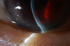
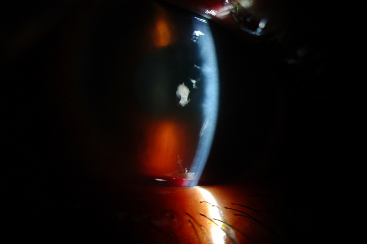
The examiner may see a taller blood level, fresh red blood over a darker clot, or new circulating cells.
However, a clot that shifts with posture can mimic a new bleed.
Therefore, record height, color and clot position at every visit (see hyphema grading).
Hyphema Rebleed Timing
Most secondary hemorrhages occur between days 2 and 5 after injury.
Also, count from the time of injury, because patients sometimes present late.
All-fit Slit-Lamp Adapter
Record and share exactly what you see at the slit lamp. One adapter fits any slit lamp or surgical microscope — and any smartphone.
From Choroida — the team behind this siteWhy the Clot Fails in That Window
Blunt trauma tears vessels in the iris or ciliary body.
Then a fibrin and platelet clot seals the damaged vessel.
Over the following days, fibrinolysis and clot retraction begin to loosen that seal.
As a result, bleeding can restart before the vessel has healed.
How Common Is a Hyphema Rebleed?
Published rates range widely, from near zero to about 38%.
Differences in populations, definitions and treatment protocols probably explain much of this spread.
Hyphema Rebleed Risk Factors
Several features recur in published series, although the strength of evidence varies.
- Larger initial hyphema
- Presenting visual acuity worse than 20/200
- Elevated IOP at presentation
- Delayed presentation after injury
- Aspirin, NSAID, antiplatelet or anticoagulant use
- Sickle cell hemoglobinopathy or another bleeding tendency
Sickle Cell Status and Bleeding History
Ask every patient about bleeding disorders, anticoagulants and family history of sickle cell disease.
Also, order hemoglobin testing when status is unknown and the patient belongs to an at-risk population.
Sickled red cells pass poorly through the trabecular meshwork, so even moderate pressure can harm the optic nerve (see sickle cell hyphema).
Monitoring a Hyphema Rebleed
Many clinicians review higher-risk patients daily through the first several days.
Each visit should include visual acuity, IOP, hyphema height, corneal clarity and a check for new blood.
However, defer gonioscopy during the bleeding window unless it would change urgent management.
Outpatient Care or Admission
Reliable patients with small hyphemas and controlled pressure can often recover at home.
On the other hand, consider admission for young children, poor adherence, large hyphemas or difficult follow-up access.
Antifibrinolytic Therapy
Aminocaproic acid and tranexamic acid slow clot breakdown, which explains their rationale.
A 2019 Cochrane review found that antifibrinolytics may reduce secondary hemorrhage.
However, the review did not show a clear gain in final visual acuity.
Aminocaproic acid can also cause nausea, vomiting and hypotension, so many clinicians reserve it for higher-risk patients.
Aspirin, NSAIDs and Anticoagulants
Advise patients to avoid aspirin and NSAIDs during recovery, and offer acetaminophen for pain.
By contrast, nobody should stop prescribed anticoagulants without weighing the systemic risk.
For example, a patient with a mechanical valve needs a joint decision with the treating physician.
Pressure After a Hyphema Rebleed
Red cells and fibrin can obstruct outflow, so pressure may climb quickly after a second bleed.
First-line options usually include a topical beta blocker or alpha agonist.
However, avoid brimonidine in young children because of the risk of systemic toxicity.
In sickle cell patients, also use carbonic anhydrase inhibitors and hyperosmotic agents with caution, because they may worsen sickling.
When Surgery Enters the Discussion
Classic thresholds for anterior chamber washout include IOP of 50 mmHg for five days or 35 mmHg for seven days.
Early corneal blood staining is another common indication.
For sickle hemoglobinopathy, surgeons often act at about 24 mmHg sustained for 24 hours.
Counseling on Hyphema Rebleed
Tell patients that the first five days carry the highest concern, even when vision seems to improve.
Also, explain that every scheduled visit matters because a pressure rise can occur silently.
Ask them to seek same-day care for any warning sign:
- Sudden drop in vision
- Worsening eye pain or headache
- Nausea or vomiting
- A visibly larger blood level
Follow-Up After the Blood Clears
Resolution of the hyphema does not end follow-up, because trauma can damage the angle and retina.
Therefore, perform gonioscopy and a dilated fundus examination once the eye is safe, often around four weeks.
Angle recession carries a long-term risk of glaucoma, so continue periodic IOP checks (see glaucoma risk factors).
Pitfalls in Hyphema Rebleed Care
- Do not discharge without a dated review plan
- Do not read a shifted clot as new bleeding without a baseline
- Never omit sickle status in at-risk patients


Document what you see
Two smartphone imaging tools built for everyday clinic use — one for the slit lamp, one for the fundus.
From Choroida — the team behind this siteReferences
- Walton W, Von Hagen S, Grigorian R, Zarbin M. Management of traumatic hyphema. Surv Ophthalmol. 2002;47:297-334.
- Gharaibeh A, Savage HI, Scherer RW, Goldberg MF, Lindsley K. Medical interventions for traumatic hyphema. Cochrane Database Syst Rev. 2019;1:CD005431.
- Crouch ER Jr, Frenkel M. Aminocaproic acid in the treatment of traumatic hyphema. Am J Ophthalmol. 1976;81:355-360.
- Deutsch TA, Weinreb RN, Goldberg MF. Indications for surgical management of hyphema in patients with sickle cell trait. Arch Ophthalmol. 1984;102:566-569.

Writes and reviews the clinical articles on Educate. Every post is checked against peer-reviewed sources before it goes live.