PVR classification grades the scar tissue that forms on and under a detached retina.
Surgeons use it to describe severity and plan repair.
Two systems dominate, the 1983 Retina Society scheme and its 1991 update.
However, neither predicts surgical success on its own.
Why PVR Classification Exists
Proliferative vitreoretinopathy develops when retinal pigment epithelial, glial and inflammatory cells form contractile membranes (see our full PVR review).
These membranes stiffen the retina and can reopen treated breaks.
Because early reports used inconsistent terms, centers could not compare results.
Therefore, surgeons needed a shared vocabulary.
The 1983 PVR Classification
The Retina Society Terminology Committee proposed four grades, A through D.
Severity increased with fold rigidity and the number of quadrants involved.
The scheme was simple, so it spread quickly.
However, it gave limited weight to anterior disease.
Fundus Explorer Pro
Photograph the retinal findings described here with the phone already in your pocket — 22 D optics and built-in illumination in one handheld unit.
From Choroida — the team behind this siteGrades A and B
Grade A shows vitreous haze and pigment clumps, which are nonspecific signs.
They can accompany a fresh break without established PVR.
Therefore, treat grade A as a warning and look for early stiffness.
Grade B adds inner retinal wrinkling, rolled break edges, vessel tortuosity and reduced mobility.
Thin membranes are often transparent, so dynamic examination helps.
Grades C and D
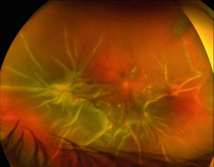
Grade C describes rigid, full thickness folds, subdivided by quadrants from C1 to C3.
Grade D means fixed folds in all four quadrants.
The retina then forms a funnel, graded wide (D1), narrow (D2) or closed (D3).
In D3, the examiner can no longer see the optic disc.
The 1991 PVR Classification Update
Machemer and colleagues kept grades A and B but redefined grade C.
The update removed grade D.
They used the equator to separate posterior disease (CP) from anterior disease (CA).
They also recorded extent in clock hours, from 1 to 12.
As a result, the report shows both location and amount of scarring.
Contraction Types in PVR Classification
The update described five contraction patterns.
First, focal contraction creates a starfold.
Next, diffuse contraction produces confluent irregular folds.
Subretinal contraction appears as a ring around the disc or as linear bands.
Circumferential contraction pulls along the posterior vitreous base, while anterior displacement drags the base forward.
Surgical Planning by PVR Classification
Anterior disease often shortens the retina and resists simple peeling.
Therefore, a CA description warns the surgeon to plan extensive vitreous base dissection.
CP disease requires peeling of posterior membranes until the retina regains mobility.
When shortening persists, a relaxing retinectomy may follow.
However, no grade dictates a single fixed operation.
Tamponade Choice and the Silicone Study
The Silicone Study randomized eyes with severe PVR to long acting tamponades.
Silicone oil gave better results than sulfur hexafluoride gas.
Meanwhile, silicone oil and perfluoropropane performed similarly overall.
Oil needs a later removal decision and can cause keratopathy, glaucoma or emulsification.
Limits of Any PVR Classification
Grading depends on the examiner and media clarity.
Intraoperative findings can also upgrade a preoperative grade.
Furthermore, the grade ignores macular status, duration and prior surgery.
Reported systems also vary between studies, which complicates comparison.
Recording the Grade in Clinic
A useful note names the system, the location and the clock hours.
For example, “CP 4, focal, inferior” says far more than “severe PVR.”
Then document findings again during surgery, because they often differ.
Examination Pearls for Early PVR
First, dilate the pupil fully and use scleral depression to view the periphery.
Next, look for a retinal surface that fails to move when the eye rotates.
Also, check the inferior retina, because membranes often start there.
In addition, note a taut, slightly shortened retina near old breaks.
Counseling Patients About Scarring
Describe PVR as scar tissue that pulls on the retina.
Explain that repeat surgery may be necessary, even after a sound operation.
Also, warn them that an attached retina does not guarantee good central vision.
Discuss positioning and oil removal.
Follow-Up After Surgery
Recurrent PVR tends to appear within the early months, so review closely.
At each visit, inspect the inferior retina, the retinectomy edge and the macula.
Also check pressure, cornea and oil position.
Adjuvant drugs remain under study, and intravitreal methotrexate is one example.
Pitfalls to Avoid
- Do not call pigment alone established PVR
- Avoid writing “grade C” without location and clock hours
- Never assume a preoperative grade covers the anterior retina
- Avoid mixing 1983 and 1991 terms in one report


Document what you see
Two smartphone imaging tools built for everyday clinic use — one for the slit lamp, one for the fundus.
From Choroida — the team behind this siteReferences
- Hilton G, Machemer R, Michels R, et al. The classification of retinal detachment with proliferative vitreoretinopathy. Ophthalmology. 1983;90:121-125.
- Machemer R, Aaberg TM, Freeman HM, et al. An updated classification of retinal detachment with proliferative vitreoretinopathy. Am J Ophthalmol. 1991;112:159-165.
- Lean JS, Stern WH, Irvine AR, Azen SP. Classification of proliferative vitreoretinopathy used in the Silicone Study. Ophthalmology. 1989;96:765-771.
- Silicone Study Group. Vitrectomy with silicone oil or sulfur hexafluoride gas in eyes with severe proliferative vitreoretinopathy: Silicone Study report 1. Arch Ophthalmol. 1992;110:770-779.
- Silicone Study Group. Vitrectomy with silicone oil or perfluoropropane gas in eyes with severe proliferative vitreoretinopathy: Silicone Study report 2. Arch Ophthalmol. 1992;110:780-789.

Writes and reviews the clinical articles on Educate. Every post is checked against peer-reviewed sources before it goes live.