Retinal emboli are particles lodged in retinal arterioles, usually from the carotid arteries, aorta or heart.
Three types account for most cases: cholesterol, platelet-fibrin and calcific.
Each has a typical appearance and, therefore, a likely source.
Why Retinal Emboli Need a Source Search
An embolus marks a proximal lesion that may release further material.
As a result, the finding can signal stroke risk and wider vascular disease.
For example, the Beaver Dam Eye Study linked retinal emboli with higher stroke-related mortality.
However, the plaque alone cannot identify the vessel or valve that released it.
Cholesterol Retinal Emboli
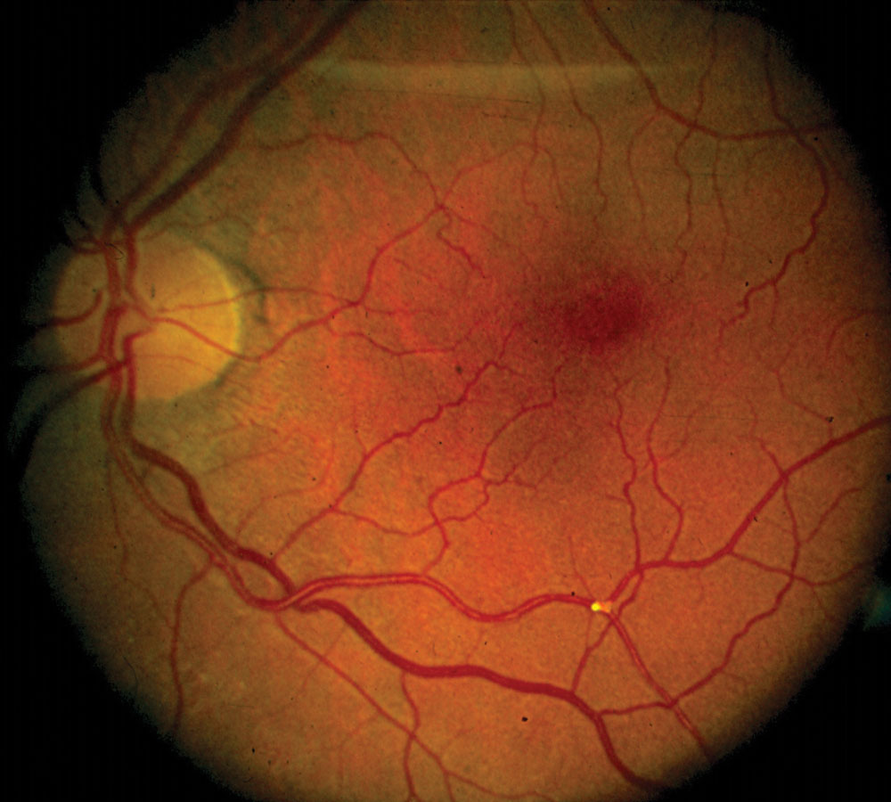
Cholesterol emboli, or Hollenhorst plaques, look bright, yellow and refractile (see Hollenhorst plaques).
They often sit at arteriolar bifurcations and usually arise from carotid or aortic atheroma.
Because they are small, blood often flows around them, so many patients report no symptoms.
Fundus Explorer Pro
Photograph the retinal findings described here with the phone already in your pocket — 22 D optics and built-in illumination in one handheld unit.
From Choroida — the team behind this siteWorkup for Cholesterol Emboli
First, check blood pressure, lipids, glucose or HbA1c, and smoking history.
Next, image the carotids with duplex ultrasound, CT angiography or MR angiography.
If the carotids look clean, consider assessing the aortic arch.
Platelet-Fibrin Retinal Emboli
These emboli look dull, gray-white and elongated, and they may fill a long arteriolar segment.
They usually form on atherosclerotic plaque or within the heart.
Because they are soft, they can break up and move downstream.
Therefore, patients often describe transient monocular vision loss, and the fundus may look normal.
Workup for Platelet-Fibrin Emboli
Treat this presentation as a possible transient ischemic attack and arrange urgent stroke evaluation.
Testing typically includes brain MRI with diffusion imaging, vessel imaging and an ECG with rhythm monitoring.
Echocardiography then looks for cardiac thrombus and other cardiac sources.
Calcific Retinal Emboli
Calcific emboli look chalky white and nonrefractile, and they are usually larger than cholesterol plaques.
They often lodge on or near the optic disc.
Because they are bulky and firm, they tend to obstruct flow and remain in place.
As a result, patients often present with a persistent central or branch retinal artery occlusion.
Workup for Calcific Emboli
Calcified aortic or mitral valve disease is the classic source, so echocardiography leads the workup.
Start with a transthoracic study, then consider a transesophageal study when suspicion stays high.
Finally, fever or a new murmur should prompt blood cultures and assessment for endocarditis.
Comparing the Three Types
- Cholesterol: bright and refractile, often asymptomatic, usually carotid or aortic
- Platelet-fibrin: dull and elongated, often transient symptoms, carotid or cardiac
- Calcific: chalky white and nonrefractile, often persistent occlusion, commonly valvular
Less Common Retinal Emboli
For example, intravenous drug use raises concern for talc, which appears as tiny refractile perimacular deposits.
Similarly, Roth spots and systemic illness may suggest septic emboli from endocarditis.
Atrial myxoma also deserves consideration in a younger patient with unexplained emboli.
Examining for Retinal Emboli
Dilate the pupil and follow each arteriole from the disc toward the periphery.
Then inspect the bifurcations closely, because small plaques often lodge there.
However, arteriolar sclerosis and vessel wall calcification can mimic an embolus, so look for an intraluminal lesion.
Imaging Retinal Emboli
Fundus photography documents the location and provides a baseline for later comparison.
OCT through the vessel may show a hyperreflective intraluminal lesion.
Also, acute ischemia produces inner retinal hyperreflectivity and swelling on OCT.
Fluorescein angiography shows delayed or absent filling.
Acute Symptoms Change the Timeline
Sudden vision loss or a recent transient episode calls for same-day evaluation, regardless of embolus type.
Current guidance treats symptomatic retinal ischemia as a stroke equivalent.
Therefore, send these patients to a stroke-capable center (see retinal artery occlusion workup).
Also consider giant cell arteritis in older patients without a visible embolus.
Asymptomatic Retinal Emboli
An incidental plaque in a patient without symptoms usually allows prompt outpatient assessment.
However, ask directly about transient vision loss, weakness, numbness and speech disturbance.
Any recent symptom should move the patient onto the urgent pathway.
Treating the Source
Treatment targets the source and aims to prevent further events.
Options include antiplatelet therapy, statins, blood pressure control and smoking cessation, while atrial fibrillation may require anticoagulation.
Carotid endarterectomy or stenting can help selected patients with symptomatic high-grade stenosis.
However, none of these measures reliably restores lost vision.
Counseling Patients With Retinal Emboli
Explain that the plaque itself may cause no visual problem, although it can signal vascular disease elsewhere.
Then advise the patient to complete every recommended test, even when vision feels normal.
Finally, teach them to call emergency services for sudden vision loss, facial droop, weakness or speech difficulty.
Pitfalls to Avoid
- Do not dismiss a plaque because vision is normal
- Do not assume a normal carotid study ends the search
- Never delay referral after recent transient monocular vision loss


Document what you see
Two smartphone imaging tools built for everyday clinic use — one for the slit lamp, one for the fundus.
From Choroida — the team behind this siteReferences
- Hollenhorst RW. Significance of bright plaques in the retinal arterioles. JAMA. 1961;178:23-29.
- Arruga J, Sanders MD. Ophthalmologic findings in 70 patients with evidence of retinal embolism. Ophthalmology. 1982;89:1336-1347.
- Klein R, Klein BE, Jensen SC, et al. Retinal emboli and stroke: the Beaver Dam Eye Study. Arch Ophthalmol. 1999;117:1063-1068.
- Biousse V, Nahab F, Newman NJ. Management of acute retinal ischemia: follow the guidelines! Ophthalmology. 2018;125:1597-1607.
- Flaxel CJ, Adelman RA, Bailey ST, et al. Retinal and ophthalmic artery occlusions Preferred Practice Pattern. Ophthalmology. 2020;127:P259-P287.

Writes and reviews the clinical articles on Educate. Every post is checked against peer-reviewed sources before it goes live.