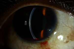
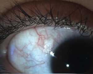
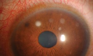
Sectoral iris atrophy is a focal loss of iris stroma and pigment epithelium, often left by herpetic uveitis.
It shows best on retroillumination, where the red reflex shines through the damaged sector.
Therefore, a careful look at transillumination can point toward the virus before any laboratory result returns.
What Sectoral Iris Atrophy Looks Like
The defect usually spans one quadrant or a wedge running from the pupil toward the iris root.
On direct illumination, the stroma looks thin, flat and pale.
In addition, the pupil margin may appear distorted or peaked toward the atrophic sector.
Why the Iris Loses Tissue
Herpesviruses inflame the iris stroma and its blood vessels.
In varicella zoster virus (VZV) disease, an occlusive vasculitis causes sectoral ischemia, then necrosis of iris tissue.
Herpes simplex virus (HSV) appears to cause more direct stromal and epithelial damage.
As a result, the pigment epithelium thins and light passes through the iris.
All-fit Slit-Lamp Adapter
Record and share exactly what you see at the slit lamp. One adapter fits any slit lamp or surgical microscope — and any smartphone.
From Choroida — the team behind this siteSectoral Iris Atrophy in VZV Uveitis
VZV uveitis often follows herpes zoster ophthalmicus, sometimes weeks after the rash.
However, some cases occur without any rash, so always ask about prior shingles.
The atrophy tends to be sectoral, sharply bordered and occasionally extensive.
Patchy Atrophy in HSV Uveitis
HSV uveitis affects younger adults more often and may follow earlier keratitis.
Its atrophy is classically patchy or moth-eaten, with scattered defects.
Also, a sectoral pattern still occurs, so shape alone cannot separate the two viruses.
Van der Lelij and colleagues described herpetic uveitis with sectoral atrophy even without keratitis.
How CMV and Fuchs Differ
Cytomegalovirus (CMV) anterior uveitis usually produces diffuse or patchy stromal atrophy with recurrent pressure spikes.
Fuchs uveitis gives diffuse thinning and heterochromia, typically without synechiae (see Fuchs heterochromic iridocyclitis).
Therefore, a well-defined wedge leans toward HSV or VZV.
Diffuse atrophy in a hypertensive eye, by contrast, raises suspicion of CMV.
Transillumination Technique
Place a narrow, short slit beam coaxially through the undilated pupil.
Next, focus on the iris plane and look for red glow through the stroma.
Dilation can hide subtle defects, so examine before drops whenever possible.
For a broader list of causes, see iris transillumination defects.
Reading the Defect Pattern
Map the defect by clock hours and photograph it at baseline.
A single sharp wedge favors VZV, while multiple scattered holes favor HSV.
Bilateral defects argue against herpes, because herpetic uveitis is almost always unilateral.
Slit Lamp Signs Around Sectoral Iris Atrophy
Herpetic uveitis commonly raises intraocular pressure through trabeculitis.
Keratic precipitates may be fine, stellate or granulomatous (see keratic precipitate patterns).
In addition, check corneal sensation and look for stromal scars.
Unilateral hypertensive uveitis with atrophy makes a strong herpetic picture.
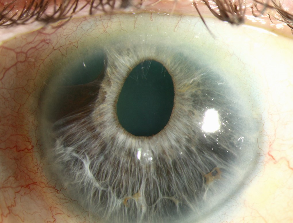
Differential Diagnosis of Sectoral Iris Atrophy
- Previous acute angle closure, which leaves ischemic atrophy and a fixed pupil
- Blunt trauma or intraocular surgery
- Pigment dispersion syndrome with bilateral radial slits
- Pseudoexfoliation with peripupillary defects
- Iridocorneal endothelial syndrome with corectopia and holes
When to Tap the Anterior Chamber
Aqueous PCR confirms the virus when the clinical picture is unclear.
A positive CMV result changes the drug class, because acyclovir has little activity against CMV.
However, a negative PCR does not exclude herpes, particularly late in the course.
Antiviral Treatment Guided by Sectoral Iris Atrophy
Sectoral atrophy in a unilateral hypertensive uveitis justifies empirical oral acyclovir, valacyclovir or famciclovir.
Published regimens use higher doses for VZV than for HSV, and renal function must guide dosing.
The HEDS iridocyclitis trial suggested benefit from adding oral acyclovir to steroids, although it was underpowered.
Later, low-dose acyclovir prophylaxis reduced recurrent ocular HSV disease in the HEDS prevention trial.
Antivirals cannot reverse established atrophy, so explain that the goal is controlling inflammation.
Steroids and Pressure Control
Topical corticosteroids control inflammation and are usually tapered slowly.
Rapid withdrawal often triggers rebound, especially in VZV disease.
Aqueous suppressants then treat elevated pressure.
Many clinicians avoid prostaglandin analogs during active herpetic inflammation, although evidence is limited.
Counseling Patients With Sectoral Iris Atrophy
Explain that the patch is a scar and will persist.
Tinted lenses or cosmetic contact lenses can reduce glare.
Also, warn that recurrences are common and that pressure may rise without symptoms.
Ask patients to return early for redness, pain or blur.
Follow-Up in Sectoral Iris Atrophy
Check pressure, cell grade and the atrophic sector at each visit.
Gonioscopy detects peripheral anterior synechiae and trabecular damage.
Next, monitor the optic nerve, because recurrent pressure spikes can cause glaucoma.
Finally, compare transillumination photos to detect enlargement.
Pitfalls to Avoid
- Do not dilate before checking transillumination
- Never stop steroids abruptly in herpetic uveitis
- Do not assume acyclovir covers CMV
- Avoid labeling bilateral defects as herpetic
- Do not miss prior angle closure as the cause
For the latest studies, see the PubMed results for sectoral iris atrophy.


Document what you see
Two smartphone imaging tools built for everyday clinic use — one for the slit lamp, one for the fundus.
From Choroida — the team behind this siteReferences
- Van der Lelij A, Ooijman FM, Kijlstra A, Rothova A. Anterior uveitis with sectoral iris atrophy in the absence of keratitis: a distinct clinical entity among herpetic eye diseases. Ophthalmology. 2000;107:1164-1170.
- Herpetic Eye Disease Study Group. A controlled trial of oral acyclovir for iridocyclitis caused by herpes simplex virus. Arch Ophthalmol. 1996;114:1065-1072.
- Herpetic Eye Disease Study Group. Acyclovir for the prevention of recurrent herpes simplex virus eye disease. N Engl J Med. 1998;339:300-306.
- Chee SP, Bacsal K, Jap A, et al. Clinical features of cytomegalovirus anterior uveitis in immunocompetent patients. Am J Ophthalmol. 2008;145:834-840.
- Liesegang TJ. Herpes zoster ophthalmicus natural history, risk factors, clinical presentation, and morbidity. Ophthalmology. 2008;115(2 Suppl):S3-S12.

Writes and reviews the clinical articles on Educate. Every post is checked against peer-reviewed sources before it goes live.