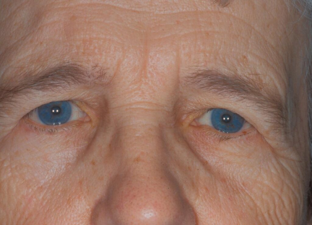
Brow ptosis is descent of the eyebrow below its normal position, usually at or below the superior orbital rim.
The drooping brow pushes forehead skin down onto the upper eyelid.
As a result, patients describe heavy lids even when the lid margin sits normally.
Recognizing the brow as the source changes the surgical plan.
Normal Brow Position
In women, the brow usually sits above the orbital rim with its peak near the lateral limbus.
In men, it lies flatter and closer to the rim.
Therefore, define a low brow against the patient’s sex, age and earlier photographs.
Why Brow Ptosis Starts Laterally
The frontalis muscle elevates the brow, but its fibers end near the temporal fusion line.
Lateral to that line, gravity and the orbicularis act with little opposition.
As a result, the tail of the brow falls first and most.
This temporal descent creates lateral hooding and a tired appearance.
All-fit Slit-Lamp Adapter
Record and share exactly what you see at the slit lamp. One adapter fits any slit lamp or surgical microscope — and any smartphone.
From Choroida — the team behind this siteCauses of Brow Ptosis
- Involutional tissue laxity with age
- Facial nerve palsy, including Bell palsy and surgical injury
- Trauma or scarring of the forehead
- Myopathies with weak frontalis, such as myotonic dystrophy
- Prior blepharoplasty that removed too much skin
- Loss of compensation after correction of true ptosis
Symptoms Patients Report
Patients complain of heavy, hooded lids and difficulty with peripheral vision.
Many notice forehead fatigue or headache at the end of the day.
Others report that lifting the brow with a finger clears their view.
Exam Signs of Brow Ptosis
First, observe the face at rest with the eyes in primary gaze.
Then look for horizontal forehead creases, which signal chronic frontalis overaction.
Next, ask the patient to close the eyes and relax the forehead.
When the eyes open again, the brow often drops to its true resting level.
Measure brow height above the pupil or the rim and record any asymmetry.
The Manual Brow Elevation Test
Lift the brow gently to a normal position with your thumb.
Then reassess the upper lid fold and the hooding.
If most of the redundant skin disappears, brow descent drives the problem.
However, persistent skin excess means dermatochalasis also needs treatment.
Brow Ptosis Versus Dermatochalasis
Dermatochalasis is redundant upper lid skin with a normally positioned brow (see dermatochalasis).
Both cause hooding, and they frequently coexist in older patients.
Because the brow supplies lid skin, a low brow mimics skin excess.
Therefore, examine the brow before you mark skin for excision.
Separating Brow Ptosis From True Ptosis
True blepharoptosis means a low lid margin, measured with the margin reflex distance (MRD1).
Brow ptosis leaves MRD1 normal once you lift the overhanging skin.
In addition, frontalis recruitment can mask both conditions.
After ptosis repair, the patient may relax the forehead, and the brow then falls (see ptosis).
Visual Field Testing
Superior visual field testing documents functional loss for insurers.
Perform it with the brow at rest and again with the brow taped up.
Improvement with taping supports a functional indication.
Blepharoplasty Alone in Brow Ptosis
Upper blepharoplasty without brow support can worsen the brow position.
Excising skin shortens the distance between brow and lashes.
Therefore, address the brow first or combine both procedures.
Direct Brow Lift
A direct brow lift excises an ellipse of skin just above the brow hairs.
It gives a powerful, predictable lift with a short operating time.
However, the scar can be visible, especially in fair, smooth skin.
It suits men with thick brows, older patients and facial palsy.
Endoscopic, Coronal and Internal Lifts
Endoscopic lifts use small incisions behind the hairline to release and fix the brow.
They hide scars well but need specific equipment and training.
Coronal lifts give strong elevation, while they risk scalp numbness, alopecia and a higher hairline.
Internal browpexy through the blepharoplasty incision stabilizes the brow, although it gives limited elevation.
Botulinum Toxin for Brow Ptosis
Botulinum toxin injected into the lateral orbicularis can raise the brow tail slightly.
This chemical brow lift suits mild cosmetic cases.
Its effect wears off after a few months.
Also, it cannot correct functional hooding that blocks the visual field.
Brow Ptosis in Facial Nerve Palsy
Paralytic brow ptosis can be marked and asymmetric.
A direct brow lift corrects it reliably.
However, check eyelid closure and the cornea before any upper face surgery.
Surgical Risks and Counseling
Supraorbital and supratrochlear nerve injury causes forehead numbness, usually temporary.
Frontal branch injury of the facial nerve can paralyze the brow.
In addition, asymmetry, recurrence and scarring can occur.
Explain that surgery repositions the brow, while fine lines and lid skin excess may need separate treatment.
Pitfalls to Avoid
- Do not perform blepharoplasty before you assess the brow
- Never measure MRD1 with skin overhanging the lid margin
- Avoid overelevating the medial brow, because it gives a surprised look


Document what you see
Two smartphone imaging tools built for everyday clinic use — one for the slit lamp, one for the fundus.
From Choroida — the team behind this siteReferences
- Booth AJ, Murray A, Tyers AG. The direct brow lift: efficacy, complications, and patient satisfaction. Br J Ophthalmol. 2004;88:688-691.
- Gunter JP, Antrobus SD. Aesthetic analysis of the eyebrows. Plast Reconstr Surg. 1997;99:1808-1816.
- American Academy of Ophthalmology. Basic and Clinical Science Course, Section 7: Oculofacial Plastic and Orbital Surgery. San Francisco: AAO; 2023.
- Salmon JF. Kanski’s Clinical Ophthalmology: A Systematic Approach. 9th ed. Elsevier; 2020.

Writes and reviews the clinical articles on Educate. Every post is checked against peer-reviewed sources before it goes live.