Uveitis testing should follow the clinical picture.
Broad batteries of tests waste money and mislead.
A targeted plan finds treatable causes.
Why Uveitis Testing Needs Selection
Many uveitis cases are idiopathic.
Tests have low yield when ordered blindly.
False positives cause confusion.
Therefore, test according to pattern and history.
Classify Before Testing
Use the SUN classification by anatomy and course.
Anterior, intermediate, posterior and panuveitis have different causes.
Acute, recurrent and chronic patterns also matter.
So define the pattern first.
All-fit Slit-Lamp Adapter
Record and share exactly what you see at the slit lamp. One adapter fits any slit lamp or surgical microscope — and any smartphone.
From Choroida — the team behind this siteUveitis Testing in a First Episode
A single mild, unilateral, nongranulomatous episode may not need tests.
Many are HLA-B27 related or idiopathic.
Observation is reasonable.
Also treat and review.
When to Test in Anterior Uveitis
Test for recurrent, bilateral, granulomatous or severe disease.
Test for associated symptoms such as back pain or rash.
Include HLA-B27 in selected patients (see HLA-B27 anterior uveitis).
So let signs drive the plan.
Syphilis in Uveitis Testing
Syphilis mimics many forms of uveitis.
Treponemal and nontreponemal tests are inexpensive.
Treatment is simple and curative.
Therefore, test any patient with unexplained uveitis.
Tuberculosis in Uveitis Testing
TB can cause granulomatous, posterior and panuveitis.
Interferon-gamma release assays help in endemic regions.
Chest imaging adds information.
Also consider it before long-term immunosuppression.
Sarcoidosis in Uveitis Testing
Chest imaging is the first step (see sarcoid uveitis).
Serum ACE and lysozyme have limited sensitivity.
Biopsy confirms the diagnosis.
So match the test to suspicion.
Uveitis Testing in Children
Juvenile idiopathic arthritis is the commonest systemic cause.
ANA testing is useful.
Regular screening of at-risk children is essential.
Therefore, work with a pediatric rheumatologist.
Posterior Uveitis
Toxoplasmosis, CMV and other infections should be considered.
Imaging with OCT and angiography helps.
Aqueous or vitreous sampling is sometimes needed.
Also check for masquerade syndromes.
Intermediate Uveitis
Pars planitis and multiple sclerosis are associated.
Sarcoidosis and tuberculosis are also causes.
MRI may be advised in selected cases.
So look beyond the eye.
Uveitis Testing and Imaging
OCT detects macular edema.
Fluorescein angiography shows vasculitis.
Fundus autofluorescence and OCT angiography help in selected diseases.
Therefore, imaging complements laboratory work.
Interpreting Results
Positive HLA-B27 does not prove cause.
Many healthy people carry it.
Interpret with clinical findings.
So avoid overdiagnosis.
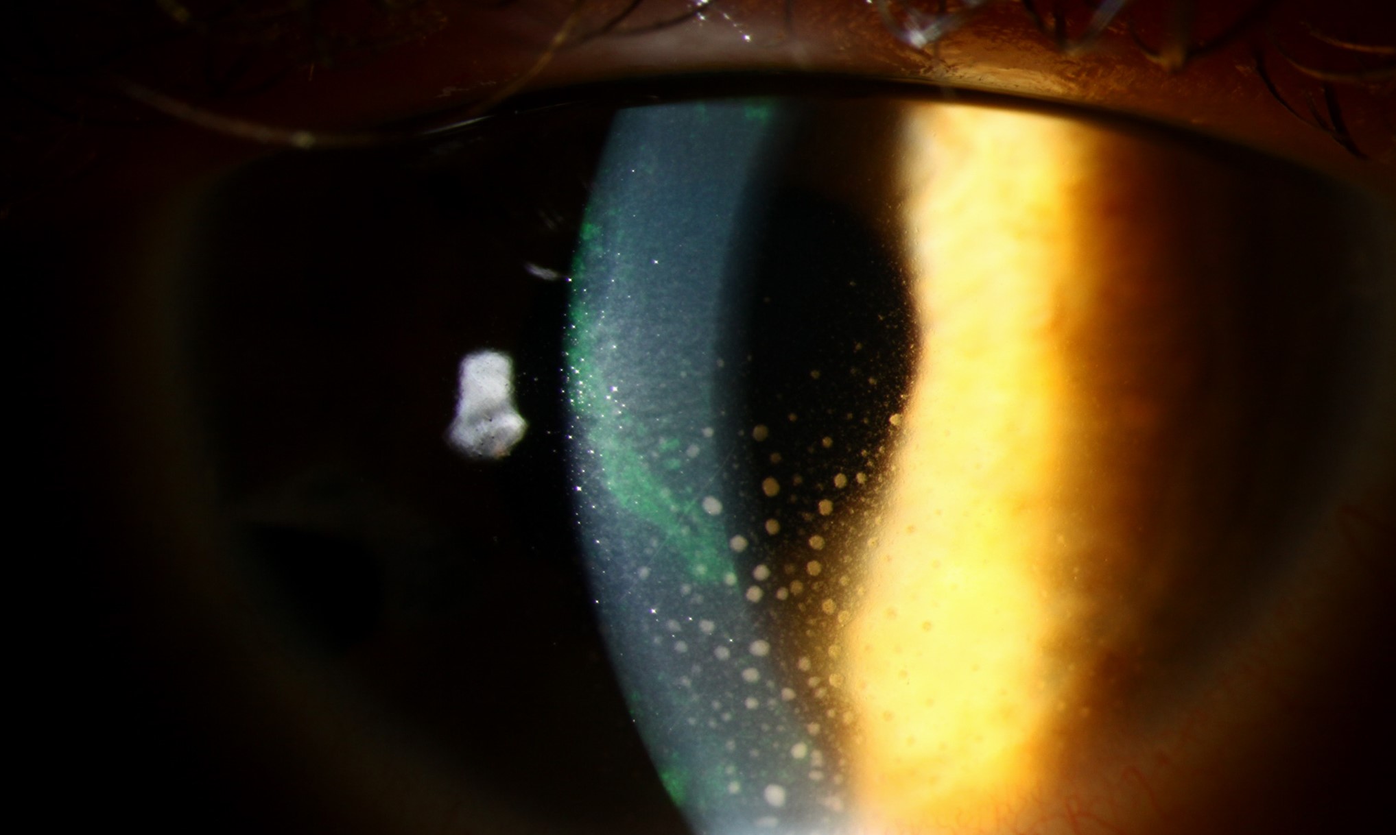
Timing and Cost
Order tests at the first visit when the pattern is clear.
Repeat only when new clues arise.
Cost matters in many health systems.
Also communicate the reasoning to patients.
Uveitis Testing: Practical Summary
Classify the uveitis, take a focused history and test selectively.
If the pattern is atypical, widen the search.
Because some causes are treatable, do not miss syphilis and TB.
Finally, coordinate with a rheumatologist when needed.
Pitfalls to Avoid
- Do not order every test for every patient
- Never ignore syphilis
- Avoid interpreting HLA-B27 in isolation
- Do not forget to screen children with arthritis
Uveitis Testing and Ankylosing Spondylitis
Back pain in the morning suggests spondyloarthritis.
HLA-B27 supports the diagnosis.
Because rheumatologists can offer treatment, refer early.
Therefore, ask about back pain and joint swelling.
Uveitis Testing and Inflammatory Bowel Disease
Bowel symptoms may accompany uveitis.
Ask about diarrhea and weight loss.
Because treatment of bowel disease can help the eye, coordinate care.
So involve the gastroenterologist.
Uveitis Testing and Masquerade Syndromes
Lymphoma and retinoblastoma can mimic uveitis.
Persistent cells in older adults raise suspicion.
Because delay is dangerous, sample the vitreous if needed.
Therefore, keep masquerade syndromes in mind.
Uveitis Testing Before Immunosuppression
Test for TB and hepatitis before starting biologics.
Check blood counts and liver function.
Because these drugs carry risks, screen carefully.
So plan tests in advance.
Uveitis Testing in Pregnancy
Toxoplasmosis and syphilis need special care.
Avoid drugs that harm the fetus.
Because many tests are safe, choose wisely.
Then coordinate with obstetrics.
Uveitis Testing: Follow-Up
Review results with the patient.
If tests are negative, continue clinical monitoring.
Because new clues can appear, repeat the history.
Finally, document the plan.
Uveitis Testing: Patient Counseling
Explain why only selected tests are ordered.
If results are negative, reassure the patient.
Because many uveitis cases are idiopathic, set realistic expectations.
Therefore, focus on controlling inflammation.
Uveitis Testing: Key Message
Classify the pattern, think of treatable causes and test with purpose.
If the picture is unusual, widen the search.
Because syphilis and TB are treatable, always consider them.
Finally, coordinate with colleagues in other specialties.
For the latest studies, see the PubMed results for uveitis testing.


Document what you see
Two smartphone imaging tools built for everyday clinic use — one for the slit lamp, one for the fundus.
From Choroida — the team behind this siteReferences
- Jabs DA, Nussenblatt RB, Rosenbaum JT; Standardization of Uveitis Nomenclature Working Group. Standardization of uveitis nomenclature for reporting clinical data. Am J Ophthalmol. 2005;140:509-516.
- Tsirouki T, Dastiridou A, Symeonidis C, et al. A focus on the epidemiology of uveitis. Ocul Immunol Inflamm. 2018;26:2-16.
- Harthan JS, Opitz DL, Fromstein SR, Morettin CE. Diagnosis and treatment of anterior uveitis: optometric management. Clin Optom. 2016;8:23-35.
- Rosenbaum JT, Bodaghi B, Couto C, et al. New observations and emerging ideas in diagnosis and management of non-infectious uveitis: a review. Semin Arthritis Rheum. 2019;49:438-445.

Writes and reviews the clinical articles on Educate. Every post is checked against peer-reviewed sources before it goes live.