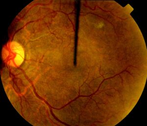
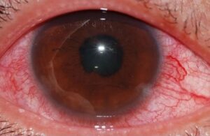
Sudden vision loss is one of the most urgent presentations in eye care.
Some causes are treatable only within hours.
A structured approach prevents missed diagnoses.
First Questions
Ask whether one eye or both are affected.
Next, ask whether the loss is painful or painless.
Finally, ask how long it has lasted.
Therefore, three questions narrow the differential at once.
Monocular vs Binocular Loss
Loss in one eye points to the eye or optic nerve.
Loss in both eyes in the same field points to the brain.
Cover each eye to confirm.
So test each eye separately.
Fundus Explorer Pro
Photograph the retinal findings described here with the phone already in your pocket — 22 D optics and built-in illumination in one handheld unit.
From Choroida — the team behind this sitePainless Sudden Vision Loss
Retinal artery occlusion, vein occlusion, detachment and vitreous hemorrhage are common causes.
Ischemic optic neuropathy also presents this way.
Wet AMD can cause sudden loss.
Also consider stroke.
Painful Sudden Vision Loss
Optic neuritis causes pain on eye movement.
Acute angle closure causes severe pain and halos.
Corneal disease and uveitis also hurt.
Therefore, pain narrows the list.
Central Retinal Artery Occlusion
Vision drops suddenly to counting fingers or worse.
The retina turns pale with a cherry-red spot (see central retinal artery occlusion).
It is a stroke equivalent.
So send the patient for urgent stroke evaluation.
Giant Cell Arteritis
Age over 50 and headache raise suspicion.
Jaw pain, scalp tenderness and weight loss add to it.
Check ESR and CRP and start steroids at once if suspected.
Also arrange temporal artery biopsy.
Central Retinal Vein Occlusion
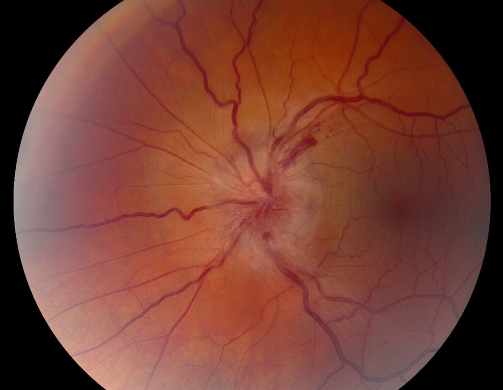
Hemorrhages cover the retina in a classic pattern.
Vision loss is usually painless and variable (see central retinal vein occlusion).
Systemic risk factors need review.
Therefore, check blood pressure and glucose.
Retinal Detachment
Flashes, floaters and a curtain are typical.
A dilated exam confirms the diagnosis.
Surgery is required.
So refer the same day (see retinal detachment symptoms and triage).
Optic Neuritis
Young adults present with pain and blurred vision.
Color vision is reduced.
MRI helps with prognosis.
Also consider multiple sclerosis.
Acute Angle Closure
The eye is red and the cornea hazy.
The pupil is mid-dilated.
Pressure is very high.
Therefore, start treatment immediately.
Transient Episodes
Brief monocular loss suggests carotid disease or arteritis (see amaurosis fugax).
Brief bilateral blur may be migraine.
Treat transient episodes as warnings.
So arrange urgent work-up.
Examination Priorities
Check visual acuity, pupils, visual fields and fundus.
Look for a relative afferent pupillary defect.
Dilate unless angle closure is suspected.
Also take blood pressure.
Investigations
OCT, fluorescein angiography and imaging of the brain may be needed.
Blood tests screen for inflammation and clotting disorders.
Choose tests by the leading diagnosis.
Therefore, avoid a blind battery of tests.
Sudden Vision Loss: Practical Summary
Triage with three questions, examine quickly and treat the dangerous causes first.
If arteritis is possible, give steroids early.
Because stroke may follow, arrange vascular work-up.
Finally, document timing carefully.
Pitfalls to Avoid
- Do not delay steroids in suspected giant cell arteritis
- Never treat artery occlusion as a routine retinal problem
- Avoid dilating a possible angle closure
- Do not forget the brain
Sudden Vision Loss in Children
Children rarely report vision loss.
Parents notice squinting or bumping into objects.
Because causes include trauma, optic neuritis and tumors, examine promptly.
Therefore, take every report seriously.
Sudden Vision Loss and Anticoagulation
Blood thinners increase the risk of vitreous hemorrhage.
Sudden dark floaters and blurred vision are typical.
Because the cause may be a tear, ultrasound may be needed.
So do not stop the medication without advice.
Sudden Vision Loss After Surgery
Endophthalmitis, detachment and macular edema can follow surgery.
Pain and redness suggest infection.
Because endophthalmitis is an emergency, refer at once.
Then start treatment without delay.
Sudden Vision Loss and Stroke
Visual loss may be the first sign of stroke.
Field defects that respect the midline suggest the brain.
Because time matters, send the patient to the emergency department.
Therefore, do not wait for outpatient imaging.
Sudden Vision Loss and Functional Causes
Some patients have no organic cause.
Examination is normal and tests do not match symptoms.
Because organic disease must be excluded first, investigate fully.
Then address the problem with sensitivity.
Sudden Vision Loss: Documentation
Record the time of onset, the best vision and the pupils.
Document the exam and the fundus.
Because later care depends on timing, be precise.
In addition, note any treatment given.
For the latest studies, see the PubMed results for sudden vision loss.


Document what you see
Two smartphone imaging tools built for everyday clinic use — one for the slit lamp, one for the fundus.
From Choroida — the team behind this siteReferences
- Mac Grory B, Schrag M, Biousse V, et al. Management of central retinal artery occlusion: a scientific statement from the American Heart Association. Stroke. 2021;52:e282-e294.
- Hayreh SS. Ocular vascular occlusive disorders: natural history of visual outcome. Prog Retin Eye Res. 2014;41:1-25.
- Hayreh SS, Zimmerman B. Visual deterioration in giant cell arteritis patients while on high doses of corticosteroid therapy. Ophthalmology. 2003;110:1204-1215.
- Pula JH, Yuen CA. Eyes and stroke: the visual aspects of cerebrovascular disease. Stroke Vasc Neurol. 2017;2:210-220.

Writes and reviews the clinical articles on Educate. Every post is checked against peer-reviewed sources before it goes live.