Systemic sclerosis is an autoimmune disease that causes skin thickening and vascular damage.
It affects many organs, including the eye.
Therefore, ocular findings can support the diagnosis and track vascular disease.
What Systemic Sclerosis Is
The disease has limited and diffuse forms.
It combines vasculopathy, immune activation and fibrosis.
Also, Raynaud’s phenomenon often comes first.
As a result, the small vessels are central to the disease.
Diagnosis of Systemic Sclerosis
The 2013 criteria of the American College of Rheumatology and EULAR score skin thickening, finger lesions and vascular signs.
Autoantibodies such as anticentromere and anti-topoisomerase help.
Also, capillaroscopy shows nailfold changes.
Therefore, the eye is one piece of a wider picture (see Sjögren syndrome).
Fundus Explorer Pro
Photograph the retinal findings described here with the phone already in your pocket — 22 D optics and built-in illumination in one handheld unit.
From Choroida — the team behind this siteEyelid Involvement in Systemic Sclerosis
Skin thickening tightens the eyelids.
Patients may have a mask-like face and reduced lid closure.
Also, telangiectasias appear on the lids and face.
As a result, exposure and dryness follow.
Ocular Surface Disease
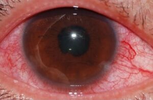
Dry eye is the most common eye complaint.
Fibrosis of the lacrimal gland and secondary Sjögren syndrome contribute.
Also, Tailor and colleagues reviewed these findings in detail.
Therefore, treat dryness early.
Retinal Vascular Changes in Systemic Sclerosis
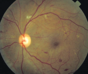
Systemic sclerosis causes narrowing and tortuosity of the retinal arterioles.
Hemorrhages and cotton-wool spots can occur.
Also, vascular occlusions are reported.
In addition, OCT angiography shows reduced vessel density in many patients.
Choroidal Involvement in Systemic Sclerosis
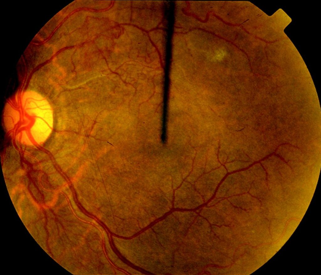
The choroid shows patchy filling defects and vascular insufficiency.
The changes mirror the microvascular damage in the skin.
Also, choroidal thinning has been reported.
Therefore, imaging can show subclinical disease.
Hypertensive Renal Crisis
Scleroderma renal crisis causes severe hypertension.
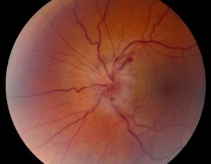
The fundus may show hypertensive retinopathy and disc swelling.
Also, the change can be sudden.
As a result, a fundus examination can warn of crisis.
Neuro-Ophthalmic Findings
Optic neuropathy and cranial nerve palsies are rare.
Giant cell arteritis may overlap in older patients.
Also, trigeminal neuropathy can cause facial pain and corneal anesthesia.
Therefore, ask about neurologic symptoms.
Drug-Related Eye Problems
Steroids raise the risk of cataract and glaucoma.
Hydroxychloroquine, when used, needs retinal screening.
Also, immunosuppressants raise the risk of opportunistic infection.
In addition, calcium channel blockers rarely cause visual symptoms.
Examination
Check the lids, ocular surface and tear film.
Dilate the pupils and examine the fundus.
Also, perform OCT and angiography when indicated.
Therefore, build a baseline for follow-up.
When to Refer
Refer patients with vision loss, red eye or new floaters.
Annual eye review is wise in diffuse disease or when taking hydroxychloroquine.
Also, urgent review is needed in a hypertensive crisis.
As a result, shared care keeps the eyes safe.
Differential Diagnosis
Other connective tissue diseases cause similar signs.
Lupus retinopathy and antiphospholipid syndrome show vascular occlusions.
Also, diabetes and hypertension cause common retinal changes.
Therefore, interpret findings within the clinical context.
Management of Eye Disease
Treat dry eye with lubricants, punctal plugs and anti-inflammatory drops.
Lid exercises and sleeping aids help with lagophthalmos.
Also, treat retinal complications as in other vascular disease.
In addition, control blood pressure strictly.
Systemic Treatment and the Eye
Immunosuppressive drugs may help skin and lung disease.
Vasodilators help Raynaud’s phenomenon and digital ulcers.
Also, ocular benefits are not well studied.
Therefore, reassess the eyes after therapy changes.
Prognosis of Systemic Sclerosis
Most patients keep useful vision.
Severe dryness and corneal complications can cause pain and scarring.
Also, retinal damage is usually mild.
As a result, regular care prevents most vision loss.
Pitfalls to Avoid
- Do not overlook eyelid tightness as a cause of exposure
- Never skip a fundus check during a hypertensive crisis
- Do not start hydroxychloroquine without a baseline retinal exam
- Never assume that retinal changes are only from hypertension
Interstitial Lung and Skin Disease
Lung fibrosis and skin thickening drive systemic treatment.
The treatment choice may affect the eyes.
Also, mycophenolate and cyclophosphamide raise infection risk.
Therefore, coordinate with the treating team.
Autoantibodies
Anticentromere antibodies link to limited disease.
Anti-topoisomerase antibodies link to diffuse disease and lung fibrosis.
Also, anti-RNA polymerase III links to renal crisis.
As a result, serology guides the risk of hypertensive eye changes.
Raynaud Phenomenon and the Eye
Vasospasm can reduce choroidal and optic nerve head flow.
Patients may report transient visual blur in cold.
Also, ocular blood flow studies show abnormal responses.
Therefore, protect patients from cold stress.
Imaging Findings
OCT angiography shows lower vessel density in the retina and choriocapillaris.
Fluorescein angiography shows slow filling in some eyes.
Also, widefield imaging may show peripheral nonperfusion.
In addition, the changes do not always cause symptoms.
For the latest studies, see the PubMed results for systemic sclerosis.


Document what you see
Two smartphone imaging tools built for everyday clinic use — one for the slit lamp, one for the fundus.
From Choroida — the team behind this siteReferences
- Tailor R, Gupta A, Herrick A, Kwartz J. Ocular manifestations of scleroderma. Surv Ophthalmol. 2009;54:292-304.
- van den Hoogen F, Khanna D, Fransen J, et al. 2013 classification criteria for systemic sclerosis: an American College of Rheumatology/European League Against Rheumatism collaborative initiative. Ann Rheum Dis. 2013;72:1747-1755.
- Denton CP, Khanna D. Systemic sclerosis. Lancet. 2017;390:1685-1699.
- Foster CS, Vitale AT. Diagnosis and Treatment of Uveitis. 2nd ed. Jaypee Brothers; 2013.

Writes and reviews the clinical articles on Educate. Every post is checked against peer-reviewed sources before it goes live.