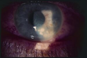
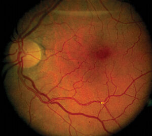
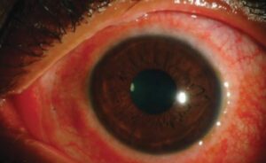
Aminoglycoside macular toxicity is damage to the macula after intraocular or periocular exposure to drugs such as gentamicin and amikacin.
It produces macular infarction, with profound and often permanent vision loss.
The condition is largely preventable, but dosing errors and dilution mistakes still occur.
What Aminoglycoside Macular Toxicity Is
Aminoglycosides are potent against gram-negative bacteria, and doctors once used them in the vitreous to treat endophthalmitis.
Even small overdoses damage the retina, particularly the macula, by causing vascular occlusion and photoreceptor loss.
Conway and Campochiaro described macular infarction after intravitreal gentamicin for endophthalmitis in the 1980s.
Campochiaro and Conway later surveyed retina specialists and documented many cases (see intracameral antibiotics).
Clinical Features of Aminoglycoside Macular Toxicity
Patients report sudden loss of vision soon after the procedure.
The fundus shows retinal whitening, hemorrhages, and cotton wool spots in the macula, and later pigment changes and atrophy.
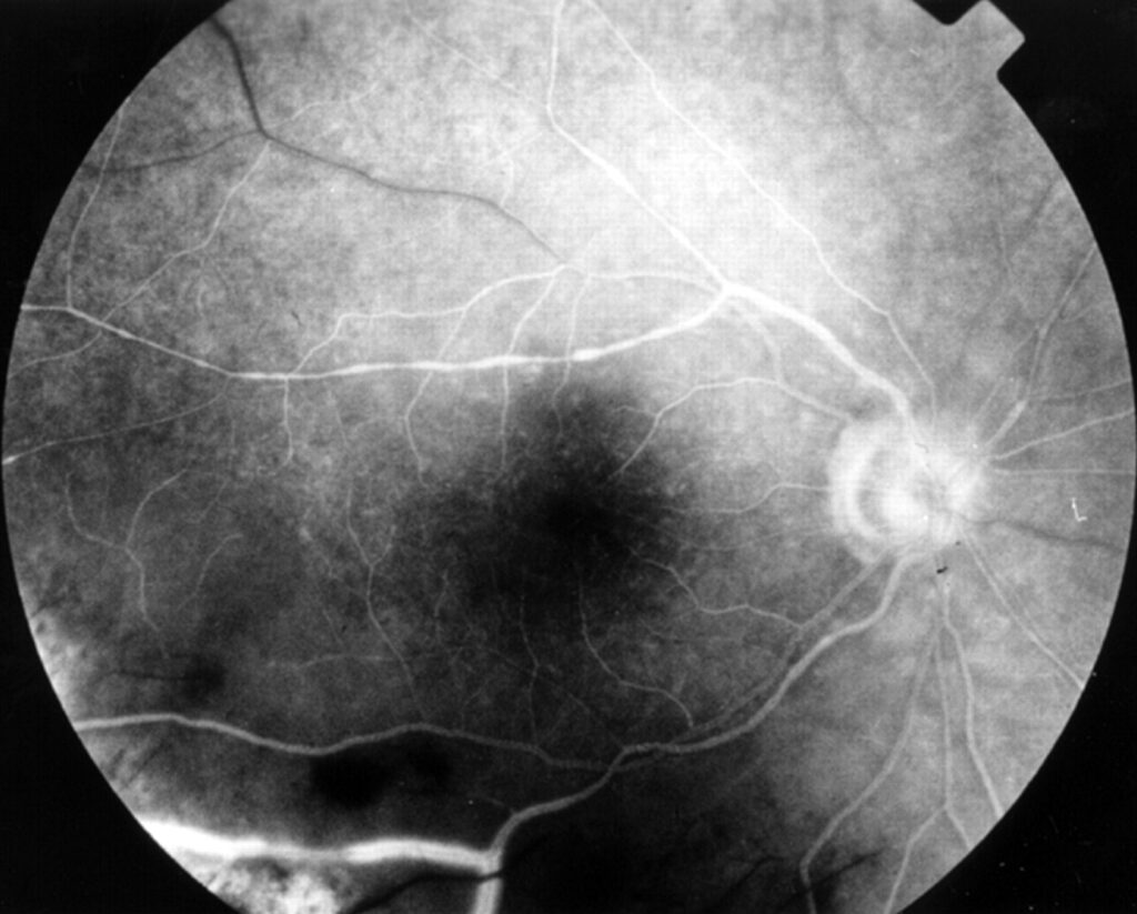
Fluorescein angiography reveals capillary nonperfusion in the macula.
OCT shows inner retinal thickening followed by atrophy.
Fundus Explorer Pro
Photograph the retinal findings described here with the phone already in your pocket — 22 D optics and built-in illumination in one handheld unit.
From Choroida — the team behind this siteDoses and Risk Factors for Aminoglycoside Macular Toxicity
The usual intravitreal gentamicin dose is about 0.1 mg, and overdoses of several times that amount are linked to macular infarction.
Amikacin carries a similar concern at excess doses, and Seawright and colleagues reported macular toxicity after intravitreal amikacin.
Risk is higher with dilution errors, repeated doses, and eyes with silicone oil or vitrectomy.
Subconjunctival and intracameral aminoglycosides can also cause damage when they leak into the eye.
How Aminoglycoside Macular Toxicity Happens in Practice
- Confusing milligram and microgram doses during dilution
- Injecting a full vial instead of a measured dose
- Using aminoglycosides in eyes with a high risk, such as diabetic eyes
- Misreading the label when preparing intravitreal antibiotics
Pharmacy preparation reduces these errors (see post-injection endophthalmitis care).
Prevention of Aminoglycoside Macular Toxicity
Prefer alternatives, such as ceftazidime, for gram-negative coverage in endophthalmitis.
Use standardized doses prepared by the pharmacy, and double-check labels and syringes.
Teach staff to report near misses, and review protocols after any event.
Management of Aminoglycoside Macular Toxicity
There is no proven treatment for established macular infarction.
Supportive care and low vision support help.
Some clinicians use hyperbaric oxygen or other measures after a recognized overdose, though evidence is limited.
Document the event and report it to the appropriate authority.
An Example Case
A 70-year-old man develops endophthalmitis after cataract surgery, and the surgeon injects intravitreal antibiotics.
A dilution error gives a much higher dose of amikacin than intended.
The next day, the retina shows macular whitening and hemorrhages, and vision falls to hand motions.
Angiography confirms capillary nonperfusion, and the macula later becomes atrophic.
The hospital reviews its protocol and moves to pharmacy-prepared syringes.
Questions Patients Ask
Patients ask whether the damage can be repaired, and the honest answer is that it usually cannot.
They also ask what went wrong, and they deserve a clear explanation and an apology when an error occurs.
Because the event has legal and safety implications, follow local reporting rules.
Pitfalls to Avoid
- Preparing antibiotic doses without a double check
- Using aminoglycosides in eyes that have already received other doses
- Ignoring symptoms of macular damage after injection
- Forgetting to review the protocol after a near miss
Looking Ahead
Standardized, pharmacy-prepared doses will lower errors.
Also, safer antibiotics offer good coverage without macular toxicity.
Therefore, aminoglycosides have lost their place in routine intravitreal therapy.
Practical Points
Use ceftazidime and vancomycin as standard intravitreal antibiotics, according to local guidelines.
Then verify each dose with a second person.
Also train staff to report errors early.
Safety Culture
Encourage reporting of near misses without blame.
Review every event to find system problems.
Because patient safety depends on small details, build checklists into the workflow.
Differential Diagnosis
Endophthalmitis itself may cause macular damage and cotton wool spots.
Therefore, ask whether the dose was correct and whether the timing fits.
Also consider retinal artery occlusion, vancomycin-associated occlusive vasculitis, and diabetic ischemia.
Because the clinical picture overlaps, review the preparation records.
Medicolegal and Safety Aspects
Disclose the event openly and document the facts.
Then report the error through hospital channels.
Also use the case to improve protocols for all staff.
Review the incident at a safety meeting within a few weeks.
Alternatives to Aminoglycosides
Ceftazidime provides gram-negative coverage with a wider safety margin.
Also vancomycin covers gram-positive organisms.
Because guidelines change, follow local protocols and consult the infectious disease team.
Then confirm doses with the pharmacy before every injection.


Document what you see
Two smartphone imaging tools built for everyday clinic use — one for the slit lamp, one for the fundus.
From Choroida — the team behind this siteReferences
- Conway BP, Campochiaro PA. Macular infarction after endophthalmitis treated with vitrectomy and intravitreal gentamicin. Arch Ophthalmol. 1986;104:367-371.
- Campochiaro PA, Conway BP. Aminoglycoside toxicity: a survey of retinal specialists. Implications for ocular use. Arch Ophthalmol. 1991;109:946-950.
- Seawright AA, Bourke RD, Cooling RJ. Macula toxicity after intravitreal amikacin. Aust N Z J Ophthalmol. 1996;24:143-146.
- American Academy of Ophthalmology. Basic and Clinical Science Course, Section 12: Retina and Vitreous. AAO; current edition.

Writes and reviews the clinical articles on Educate. Every post is checked against peer-reviewed sources before it goes live.