Proton beam therapy delivers a focused dose of charged particles to an intraocular tumor and spares most of the surrounding eye.
It treats uveal melanoma that is too large or too close to the optic disc for plaque brachytherapy.
Specialized centers offer it, and the referral decision rests on tumor size, location, and the patient’s goals.
How Proton Beam Therapy Works
In contrast to photons, protons release most of their energy at a defined depth, called the Bragg peak.
Therefore, the beam covers the tumor with a high dose and stops behind it.
Surgeons first sew small tantalum clips to the sclera around the tumor, and these clips guide the beam.
The patient then receives about four fractions over several days, with the head fixed in a mask.
Indications for Proton Beam Therapy
- Large melanomas beyond the limits of plaque therapy
- Tumors near the optic disc or fovea
- Ciliary body tumors, in selected centers
- Eyes where plaque placement is technically hard
Small tumors may still receive plaque treatment, and very large tumors may need enucleation (see plaque brachytherapy for choroidal melanoma).
Fundus Explorer Pro
Photograph the retinal findings described here with the phone already in your pocket — 22 D optics and built-in illumination in one handheld unit.
From Choroida — the team behind this siteLocal Control and Survival
Overall, large series report tumor control above 90 percent at five years.
The Collaborative Ocular Melanoma Study showed that radiation and enucleation gave similar survival for medium tumors.
Therefore, eye preservation does not shorten life for patients with medium tumors, although the risk of metastasis depends on the tumor biology (see choroidal melanoma).
Meta-analyses suggest that charged particle therapy achieves better local control than plaque therapy in some series.
Complications of Proton Beam Therapy
Radiation damage appears months to years after treatment, so follow-up must continue for life.
- Radiation retinopathy and maculopathy
- Optic neuropathy from damage to the nerve head
- Neovascular glaucoma
- Cataract and dry eye
Anti-VEGF injections can slow radiation retinopathy and edema, though they do not prevent it in every eye, and laser or steroids may help selected eyes (see radiation retinopathy).
Follow-Up After Proton Beam Therapy
Examine the eye every three to six months in the first years.
Also document tumor regression with fundus photographs and ultrasound.
Dry eye, lash loss, and eyelid changes may occur when the beam passes through the anterior structures.
Watch the macula and disc with OCT, and check the pressure and angle for neovascular changes.
Metastatic surveillance, usually with liver imaging, continues for life under the oncology team.
Refer promptly when the tumor grows or when new subretinal fluid appears after a long quiet period.
Counselling
Because vision usually declines when the tumor lies near the macula or disc, explain this early.
Tell patients that the tumor may shrink slowly over a year or more.
Describe the clip surgery and the treatment schedule in advance.
An Example Case
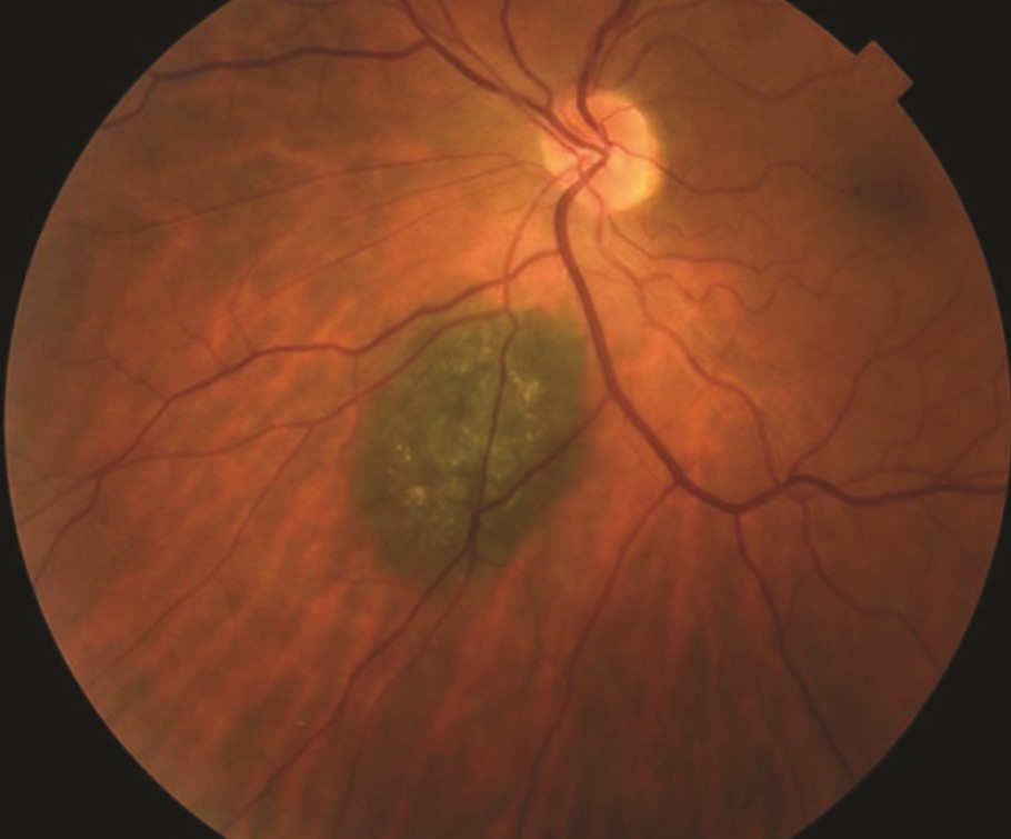
A 58-year-old man has a 9 mm wide choroidal melanoma touching the optic disc, with a thickness of 6 mm.
Plaque therapy would risk an inadequate dose near the disc.
After staging, he has tantalum clips placed in the sclera, and he receives four fractions of proton beam therapy.
The tumor shrinks over 18 months, and vision falls from 20/30 to 20/100 because of radiation optic neuropathy.
He keeps the eye, has regular liver imaging, and remains free of metastases at five years.
Proton Beam Therapy Versus Plaque Therapy
Plaque therapy is available in more centers, and it controls most small and medium tumors.
Proton beam therapy fits tumors that sit near the disc or fovea, or that are too wide for a plaque.
Therefore, refer difficult tumors early, since planning takes time.
Pitfalls to Avoid
- Refer early instead of waiting for tumor growth
- Explain the risk of late vision loss before treatment
- Continue lifelong screening for liver metastases
- Check the history before labeling radiation retinopathy as diabetic disease
Questions Patients Ask
Patients ask whether they will lose the eye, and many keep the eye after radiation.
They also ask whether treatment prevents metastasis, and the honest answer is that it controls the local tumor, not the biology.
Describe the follow-up schedule and the role of the oncology team.
Key Points
Proton beam therapy delivers a precise dose to uveal melanoma and spares nearby tissue.
It gives high local control and suits tumors near the disc or macula.
Late radiation retinopathy and optic neuropathy remain common reasons for vision loss.
For the latest studies, see the PubMed results for proton beam therapy.


Document what you see
Two smartphone imaging tools built for everyday clinic use — one for the slit lamp, one for the fundus.
From Choroida — the team behind this siteReferences
- Gragoudas ES. Proton beam irradiation of uveal melanomas: the first 30 years. The Weisenfeld Lecture. Invest Ophthalmol Vis Sci. 2006;47:4666-4673.
- Egger E, Zografos L, Schalenbourg A, et al. Eye retention after proton beam radiotherapy for uveal melanoma. Int J Radiat Oncol Biol Phys. 2003;55:867-880.
- Wang Z, Nabhan M, Schild SE, et al. Charged particle radiation therapy for uveal melanoma: a systematic review and meta-analysis. Int J Radiat Oncol Biol Phys. 2013;86:18-26.
- Collaborative Ocular Melanoma Study Group. The COMS randomized trial of iodine 125 brachytherapy for choroidal melanoma: V. Twelve-year mortality rates and prognostic factors: COMS report No. 28. Arch Ophthalmol. 2006;124:1684-1693.

Writes and reviews the clinical articles on Educate. Every post is checked against peer-reviewed sources before it goes live.