Optogenetic therapy for retinal degeneration tries to give vision back to eyes whose photoreceptors have died.
Instead of repairing a specific gene, it makes the remaining cells of the inner retina sensitive to light, so it can work in many diseases.
It is early, but it has produced the first reports of partial recovery of vision in a blind patient.
The Problem Optogenetic Therapy for Retinal Degeneration Addresses
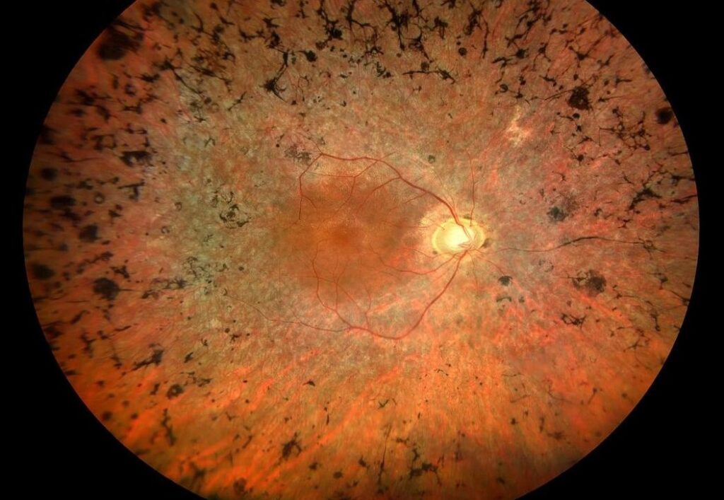
In advanced retinitis pigmentosa and some other diseases, rods and cones are lost, but many inner retinal neurons survive (see retinitis pigmentosa).
These cells still connect to the optic nerve, but they receive no light signal.
Gene therapy for a single gene helps only patients with that mutation, and it needs surviving photoreceptors (see gene therapy for inherited retinal disease).
Optogenetic Therapy for Retinal Degeneration: How Optogenetics Works
- A viral vector delivers the gene for a light-sensitive protein, such as channelrhodopsin, into surviving cells
- The cells then respond to light by firing electrical signals
- The protein is derived from algae, microbes, or human opsins, and it differs in light sensitivity and speed (see the references)
- Targets include retinal ganglion cells, bipolar cells, or remaining cone bodies
The approach is independent of the original gene defect, which is its main advantage.
Fundus Explorer Pro
Photograph the retinal findings described here with the phone already in your pocket — 22 D optics and built-in illumination in one handheld unit.
From Choroida — the team behind this siteWhat the First Results Showed
In 2021, a report described a man with advanced retinitis pigmentosa who received an intravitreal injection of a vector carrying a channelrhodopsin, followed by training with light-amplifying goggles.
He could locate and count objects with the treated eye while wearing the goggles, which he could not do before, and brain recordings showed responses to the objects (see the references).
Other studies have used different proteins and delivery methods, and some report improvements on visual function tests.
Optogenetic Therapy for Retinal Degeneration: The Role of Goggles
Optogenetic proteins are less sensitive than natural photoreceptors, so the light from the environment must be converted to intense pulses of a specific color.
The patient wears goggles with a camera, which projects the image onto the retina, and the patient must also learn to use the new signals.
Limits and Risks
- Vision is crude, and it is far from normal sight
- The light intensity needed can damage the retina if it is too strong
- Inflammation or an immune response to the vector can occur
- Results vary, and long-term durability is unknown
- Treatment is limited to patients with severe loss and surviving inner retinal cells
Optogenetic Therapy for Retinal Degeneration: Who Might Benefit?
Patients with advanced inherited retinal degeneration, with light perception or bare vision, are the first candidates.
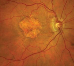
Trials in other diseases, such as geographic atrophy, are being planned.
Comparison With Other Options
- Retinal implants stimulate the retina electrically, require surgery, and give coarse vision (see retinal implants)
- Stem cell therapy tries to replace lost cells (see stem cell therapy for retinal disease)
- Mutation-specific gene therapy corrects the defect when cells are still alive
These strategies may complement each other in the future.
Optogenetic Therapy for Retinal Degeneration: Counselling
Patients should know that these treatments are experimental, that trials have strict criteria, and that unproven clinics should be avoided.
Also, give them information about registered trials and genetic testing.
Safety and Immune Response
Vectors delivered into the eye can trigger inflammation, and patients usually receive steroids around the procedure.
Intravitreal delivery is simpler than subretinal surgery, but it needs enough protein expression in the target cells, and the vitreous and inner limiting membrane can reduce transduction.
Researchers are testing engineered capsids that cross these barriers better.
Training and Rehabilitation
After treatment, patients need weeks to months of training with the device, and they learn to interpret the new signals.
Support from rehabilitation teams is part of the plan, and outcome measures include navigation, object recognition, and quality of life.
Eligibility and Trials
Eligible patients usually have light perception or hand movements, have lost reading vision, and have documented inherited disease.
Trials follow strict criteria, and patients should seek registered studies, and avoid clinics that offer cell or gene injections without oversight.
Future Directions
- More sensitive proteins that work in ambient light and do not need goggles
- Targeting of specific cell types for better resolution
- Combination with retinal prostheses or cell therapy
- Use in diseases beyond inherited degeneration
Key Messages for Colleagues
Optogenetics is the first approach that could help patients with many types of advanced blindness.
It is the most advanced among strategies that do not depend on the gene.
Clinicians can prepare by documenting the retinal findings, the visual function, and the genetic test result, so that suitable patients can be referred to trials.
For the latest studies, see the PubMed results for optogenetic therapy for retinal degeneration.


Document what you see
Two smartphone imaging tools built for everyday clinic use — one for the slit lamp, one for the fundus.
From Choroida — the team behind this siteReferences
- Sahel JA, Boulanger-Scemama E, Pagot C, et al. Partial recovery of visual function in a blind patient after optogenetic therapy. Nat Med. 2021;27:1223-1229.
- Bi A, Cui J, Ma YP, et al. Ectopic expression of a microbial-type rhodopsin restores visual responses in mice with photoreceptor degeneration. Neuron. 2006;50:23-33.
- Cehajic-Kapetanovic J, Eleftheriou C, Allen AE, et al. Restoration of vision with ectopic expression of human rod opsin. Curr Biol. 2015;25:2111-2122.
- American Academy of Ophthalmology. Basic and Clinical Science Course, Section 12: Retina and Vitreous. AAO; current edition.

Writes and reviews the clinical articles on Educate. Every post is checked against peer-reviewed sources before it goes live.