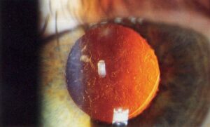
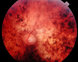
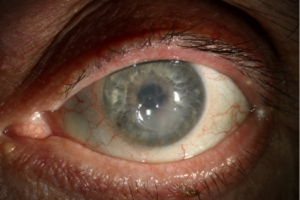
Corneal laceration repair restores the integrity of the eye after a cut through the cornea.
Also, the goals are to close the wound watertight, restore the shape of the cornea, protect the lens, and reduce astigmatism and scarring.
The approach depends on whether the wound is partial or full thickness, and on its size and shape.
Classification Before Corneal Laceration Repair
- Partial-thickness laceration: does not enter the anterior chamber, and has a negative Seidel test
- Full-thickness laceration: enters the anterior chamber, with a leak, shallow chamber, or iris prolapse
- Simple or stellate: a clean cut or a complex tear with multiple limbs
- With tissue loss or iris incarceration, which needs special care
Also, the Birmingham Eye Trauma Terminology classifies the injury as an open globe injury when the wall is penetrated (see the references).
Corneal Laceration Repair: Initial Assessment
- Examine gently, without pressure, and shield the eye (see ocular first aid)
- Check the visual acuity, the pupil, and the shape of the chamber
- Perform a Seidel test with care, and look for aqueous leak
- Exclude an intraocular foreign body and posterior rupture, with CT when needed (see ruptured globe)
- Give tetanus prophylaxis and antibiotics, and keep the patient fasting
All-fit Slit-Lamp Adapter
Record and share exactly what you see at the slit lamp. One adapter fits any slit lamp or surgical microscope — and any smartphone.
From Choroida — the team behind this siteSmall Self-Sealing Wounds
Short, oblique wounds with a formed chamber and no leak may also seal by themselves.
- A bandage contact lens, antibiotics, and close follow-up
- Cyanoacrylate glue for small leaks, up to about 2 to 3 millimeters (see corneal perforation glue)
- Watch for a flat chamber and reopening
Corneal Laceration Repair: Suturing Principles
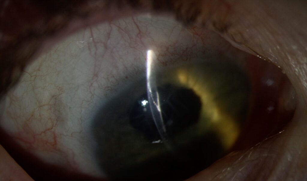
Wounds that leak, gape, or have a shallow chamber, however, need sutures.
- Use 10-0 nylon, with a spatulated needle
- Take bites at about 90 percent of the corneal thickness, on both sides of the wound, and at equal depth
- Space sutures about 1.5 to 2 millimeters apart, with bites 1 to 1.5 millimeters from the wound edge
- Start with the sutures that align landmarks, such as the limbus or a wound angle
- Bury the knots on the corneal side that is deeper, so that they do not irritate the lid
- Check for leaks with fluorescein after closure
In addition, sutures should be shorter in the center, to avoid inducing astigmatism and to keep the visual axis clear.
Dealing With Iris and Lens
- Reposition clean iris with viscoelastic, and excise only if it is dead or contaminated
- Reform the chamber with viscoelastic or balanced salt solution through a side port
- Examine the lens, and remove it if the capsule is torn with cortex in the chamber (see traumatic cataract)
- Take care not to touch the endothelium, because it does not regenerate
Stellate and Complex Wounds
- Place a central purse-string suture to bring the flaps together, or use multiple sutures at the angles
- Glue and a bandage lens can also supplement the suture
- Tissue loss may also require a patch graft or a corneal transplant
Postoperative Care
- Topical antibiotics and steroids, and a cycloplegic
- Systemic antibiotics in contaminated wounds, with consideration of intravitreal antibiotics for retained organic foreign bodies
- Monitor the pressure, the chamber depth, and the sutures
- Remove or adjust sutures as the wound heals, at three to six months, to reduce astigmatism
- Refract and consider rigid lenses or keratoplasty for visual rehabilitation (see post-keratoplasty astigmatism)
Complications of Corneal Laceration Repair
- Endophthalmitis, a major risk of open globe injury
- Traumatic cataract and lens injury
- Irregular astigmatism and scarring
- Glaucoma, retinal detachment, and proliferative vitreoretinopathy
Early referral and a coordinated plan also improve results.
Anesthesia and Setting
Repair of a full-thickness wound is usually done in the operating room under general anesthesia or a block, with care to avoid pressure on the eye.
Avoid retrobulbar blocks when an open globe is suspected, since they can push out intraocular contents.
Topical anesthesia may also be used for small partial-thickness wounds in a cooperative adult.
Common Pitfalls
- Sutures that are too tight, which cause steep astigmatism
- Sutures that are too shallow, which leak
- Missing a second wound, such as a scleral laceration or a posterior extension
- Leaving iris in the wound, which delays healing and raises the infection risk
- Failing to check for a foreign body
A careful inspection of the whole globe after the repair helps to avoid missing associated injuries.
Visual Outcomes
Visual outcome depends on the location of the wound, the involvement of the lens and the retina, and the speed of repair.
Central wounds cause more scarring, and many patients need rigid lenses or other procedures to restore vision after the wound has healed.
For the latest studies, see the PubMed results for corneal laceration repair.


Document what you see
Two smartphone imaging tools built for everyday clinic use — one for the slit lamp, one for the fundus.
From Choroida — the team behind this siteReferences
- Kuhn F, Morris R, Witherspoon CD. Birmingham Eye Trauma Terminology (BETT): terminology and classification of mechanical eye injuries. Ophthalmol Clin North Am. 2002;15:139-143.
- Pieramici DJ, Sternberg P Jr, Aaberg TM Sr, et al. A system for classifying mechanical injuries of the eye (globe). Am J Ophthalmol. 1997;123:820-831.
- Kuhn F, Pieramici DJ. Ocular Trauma: Principles and Practice. Thieme; 2002.
- American Academy of Ophthalmology. Basic and Clinical Science Course, Section 8: External Disease and Cornea. AAO; current edition.

Writes and reviews the clinical articles on Educate. Every post is checked against peer-reviewed sources before it goes live.