
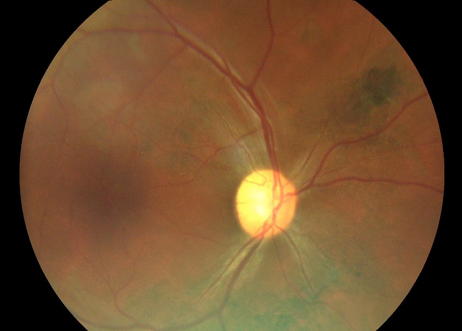
Post-Ebola uveitis is an intraocular inflammation seen in survivors of Ebola virus disease, usually in the months after recovery.
It causes visual loss and blindness in a notable proportion of survivors in the affected regions.
Viral RNA has been found in the eye after it was cleared from the blood, which changes how clinicians approach both treatment and surgery.
What Is Post-Ebola Uveitis?
The West African epidemic of 2014 to 2016 left thousands of survivors, and eye complications emerged as a major cause of morbidity.
Studies from Sierra Leone and Liberia found uveitis in a substantial fraction of survivors, with a higher risk in those who had a high viral load during the acute illness.
The eye is an immune-privileged site where the virus may persist, away from the effect of the antibody response.
A report in 2015 described Ebola virus RNA in the aqueous humor of a physician who had recovered, 14 weeks after the virus was no longer detected in blood.
Clinical Features
Patients present with redness, pain, photophobia, floaters, and reduced vision, usually in the first year after the illness.
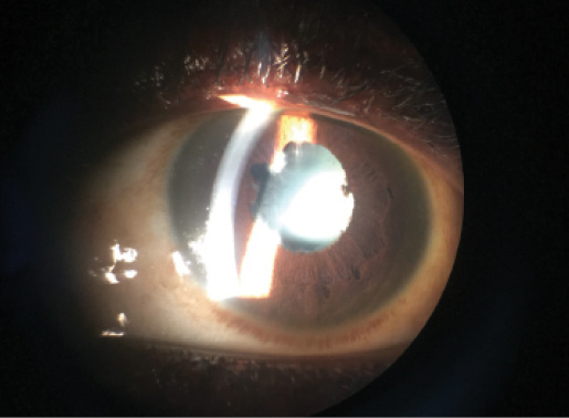
- Anterior uveitis: with synechiae, and sometimes a hypopyon
- Intermediate and posterior uveitis: vitritis, vasculitis, and chorioretinitis
- Panuveitis: the most severe form
- Cataract and glaucoma: frequent complications
- Optic neuritis and retinal scars: reported in some survivors
Posterior synechiae, raised pressure, and cataract are common causes of vision loss (see posterior synechiae).
Peripheral chorioretinal scars have been noted in survivors and may be a sign of past inflammation.
All-fit Slit-Lamp Adapter
Record and share exactly what you see at the slit lamp. One adapter fits any slit lamp or surgical microscope — and any smartphone.
From Choroida — the team behind this siteDifferential Diagnosis
- Other infectious uveitis common in West Africa: onchocerciasis, toxoplasmosis, tuberculosis, and syphilis
- Viral uveitis from herpes viruses: see viral anterior uveitis
- Acute retinal necrosis: a necrotizing retinitis (see acute retinal necrosis)
- Noninfectious uveitis: autoimmune causes
The history of Ebola virus disease, usually documented by a survivor certificate, makes the diagnosis likely.
Management
Treatment is based on corticosteroids, topical and systemic, together with cycloplegics.
The steroid response is generally good, but the safety of immunosuppression in a person with a possible persistent viral reservoir has been debated.
Survivors with uveitis have been treated with systemic steroids in studies and clinics, and the benefit appears to outweigh the risk when inflammation threatens vision.
Antiviral agents have been used in clinical programs, but their role is not established.
Monitor the pressure, treat the glaucoma, and follow the lens.
Surgery and Safety
Cataract surgery in survivors is important for restoring vision, and it has been performed safely in many cases, in programs with strict precautions.
Because viable virus may be present in aqueous humor, the surgical team should use personal protective equipment, safe handling of fluids, and careful disposal.
Surgery is planned when the eye has been quiet for some time, and the patient receives perioperative anti-inflammatory therapy.
Counsel survivors about regular eye examinations, because the problem can be silent, and about the stigma that they may meet in their communities.
Scale of the Problem
Surveys in Sierra Leone and Liberia reported uveitis in roughly one in ten to one in three survivors who were examined, and the frequency increased with time since the illness in some series.
Many survivors lost vision from cataract and glaucoma that followed the inflammation.
The need for eye care continued long after the epidemic was declared over, and ophthalmologists in the region developed special clinics.
Examination in Limited Settings
A portable slit lamp, a handheld tonometer, and an indirect ophthalmoscope are the basic tools.
Check visual acuity, pupils, the anterior chamber, the pressure, and the lens, and examine the fundus through a dilated pupil.
Document the findings, and consider ultrasound when the view is obscured.
Where PCR of aqueous humor is available, it can detect viral RNA, but treatment decisions usually rely on the clinical picture.
Counselling
Survivors often face stigma and fear of discrimination, so a respectful approach and clear communication are part of the care.
Explain that the eye disease is the consequence of the infection and not a new infection, that it can be treated, and that regular follow-up reduces the risk of blindness.
Make sure that patients understand how to use their drops and when to return.
Rehabilitation
Many survivors with visual loss need low-vision services, and support from community eye health programs helps them to return to work.
Cataract surgery programs for survivors, combined with spectacles and drops, restore sight for many, and they should be integrated with general eye care.
Share the experience with colleagues and with health authorities, because the same pattern may occur in future outbreaks.


Document what you see
Two smartphone imaging tools built for everyday clinic use — one for the slit lamp, one for the fundus.
From Choroida — the team behind this siteReferences
- Varkey JB, Shantha JG, Crozier I, et al. Persistence of Ebola virus in ocular fluid during convalescence. N Engl J Med. 2015;372:2423-2427.
- Mattia JG, Vandy MJ, Chang JC, et al. Early clinical sequelae of Ebola virus disease in Sierra Leone: a cross-sectional study. Lancet Infect Dis. 2016;16:331-338.
- Shantha JG, Crozier I, Hayek BR, et al. Ophthalmic manifestations and causes of vision impairment in Ebola virus disease survivors in Monrovia, Liberia. Ophthalmology. 2017;124:170-177.
- American Academy of Ophthalmology. Basic and Clinical Science Course, Section 9: Uveitis and Ocular Inflammation. AAO; current edition.

Writes and reviews the clinical articles on Educate. Every post is checked against peer-reviewed sources before it goes live.


