
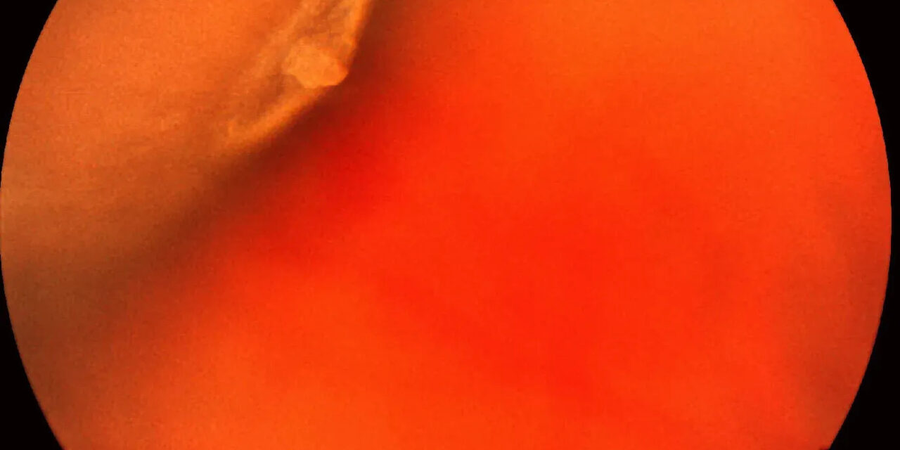
Retinal tufts are small, focal areas of firm vitreoretinal adhesion in the peripheral retina, and while any single tuft is a minor finding on its own, understanding which specific type carries meaningful risk, and, even more importantly, recognizing when new symptoms suggest that risk has become active, matters more than the finding’s small physical size might suggest.
What Retinal Tufts Are
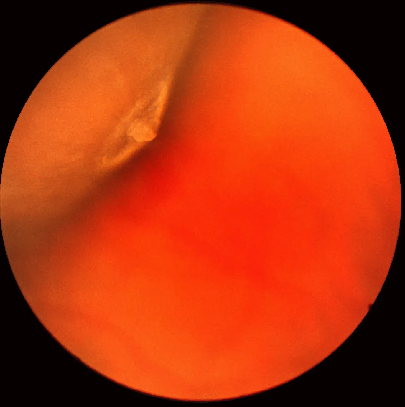
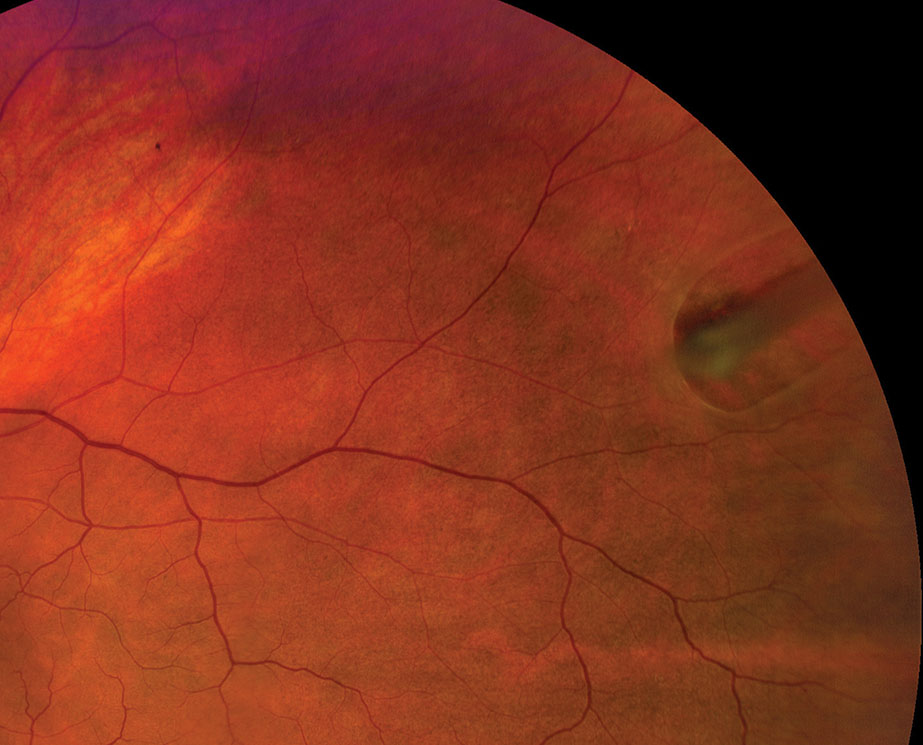
Retinal tufts represent small, focal areas where the vitreous is more firmly adherent to the peripheral retina than in the surrounding area, and this firm, focal adhesion is the mechanistic reason tufts are relevant to retinal tear risk: if the vitreous undergoes traction at this point of unusually strong adhesion, such as during a posterior vitreous detachment, the retina at that specific point can tear rather than releasing cleanly.
Classification
Noncystic Retinal Tufts
The more common type, generally small and considered to carry a very low risk of associated retinal tear or detachment in the absence of any acute symptoms, and typically managed with observation alone.
Cystic Retinal Tufts
Somewhat less common, these tufts have an associated area of cystic retinal degeneration and are generally considered to carry a modestly higher risk than noncystic tufts, given their association with a greater degree of chronic vitreoretinal traction.
Zonular Traction Tufts
A specific type located near the ora serrata, associated with traction from zonular fibers rather than purely vitreous traction, another recognized variant within this broader category of focal peripheral vitreoretinal adhesions.
Fundus Explorer Pro
Photograph the retinal findings described here with the phone already in your pocket — 22 D optics and built-in illumination in one handheld unit.
From Choroida — the team behind this siteClinical Significance
The overwhelming majority of retinal tufts, of any type, remain entirely stable throughout a patient’s lifetime and never cause a retinal tear or detachment, which is why the finding of an asymptomatic tuft on routine dilated examination is, in itself, generally not considered a sufficient reason for prophylactic treatment.
The clinical significance of a retinal tuft changes substantially, however, when a patient presents with new, acute symptoms, particularly new flashes and floaters suggestive of an acute posterior vitreous detachment, since this is precisely the scenario in which the firm adhesion at a tuft can be pulled upon and produce an actual retinal tear.
Clinical Evaluation
- Dilated fundus examination with scleral depression is needed to adequately visualize and characterize peripheral retinal tufts, since many are located in the far peripheral retina
- Careful assessment of whether a tuft is cystic or noncystic, and whether there is any associated retinal break, hemorrhage, or other sign of acute traction
- In a patient presenting with new, acute flashes and floaters, particular attention to any known or newly identified retinal tufts is warranted, given their recognized association with tear formation during acute posterior vitreous detachment
Management
Asymptomatic Tufts
Asymptomatic retinal tufts identified incidentally on routine examination, without any associated retinal break or other acute finding, are generally managed with observation and patient education about the symptoms of a posterior vitreous detachment (new flashes, a sudden increase in floaters, or a curtain-like visual field loss) that should prompt urgent reevaluation.
Symptomatic Presentation or Associated Break
When a patient presents with acute symptoms and a retinal tuft is found to be associated with an actual retinal tear, or when a tuft shows other signs of acute traction, prophylactic laser retinopexy or cryotherapy is performed around the area to create a chorioretinal adhesion and prevent subretinal fluid from tracking through any associated break, following the same general principles applied to any acute, symptomatic retinal tear.
Patient Education
Given that the risk associated with a retinal tuft is meaningfully greater during an acute posterior vitreous detachment than at baseline, education about recognizing and promptly reporting new flashes, floaters, or visual field changes is a valuable and appropriately targeted part of counseling any patient found to have this finding on routine examination.
Prognosis
The great majority of patients with incidentally discovered retinal tufts never experience any related complication and require nothing beyond routine, age-appropriate eye care.
For the smaller number who develop an acute posterior vitreous detachment with associated traction at the site of a tuft, prompt recognition of new symptoms and timely examination allows any resulting tear to be identified and treated before progression to retinal detachment, which is the entire rationale behind patient education at the time this finding is first identified.


Document what you see
Two smartphone imaging tools built for everyday clinic use — one for the slit lamp, one for the fundus.
From Choroida — the team behind this siteReferences
- Byer NE. Cystic retinal tufts and their relationship to retinal detachment. Arch Ophthalmol. 1981;99:1788-1790.
- Foos RY. Zonular traction tufts of the peripheral retina in cadaver eyes. Arch Ophthalmol. 1969;82:620-632.
- Wilkinson CP. Evidence-based analysis of prophylactic treatment of asymptomatic retinal breaks and lattice degeneration. Ophthalmology. 2000;107:12-15.
- Byer NE. What happens to untreated asymptomatic retinal breaks, and are they affected by posterior vitreous detachment? Ophthalmology. 1998;105:1045-1049.
