
Corneal guttae are small, wart-like excrescences of Descemet membrane produced by dysfunctional endothelial cells, and because they appear years, sometimes decades, before any corneal edema develops, recognizing them at the slit lamp gives a genuinely early warning of endothelial trouble ahead, well before a patient has any symptoms at all.
What Guttae Actually Are
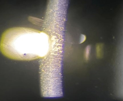
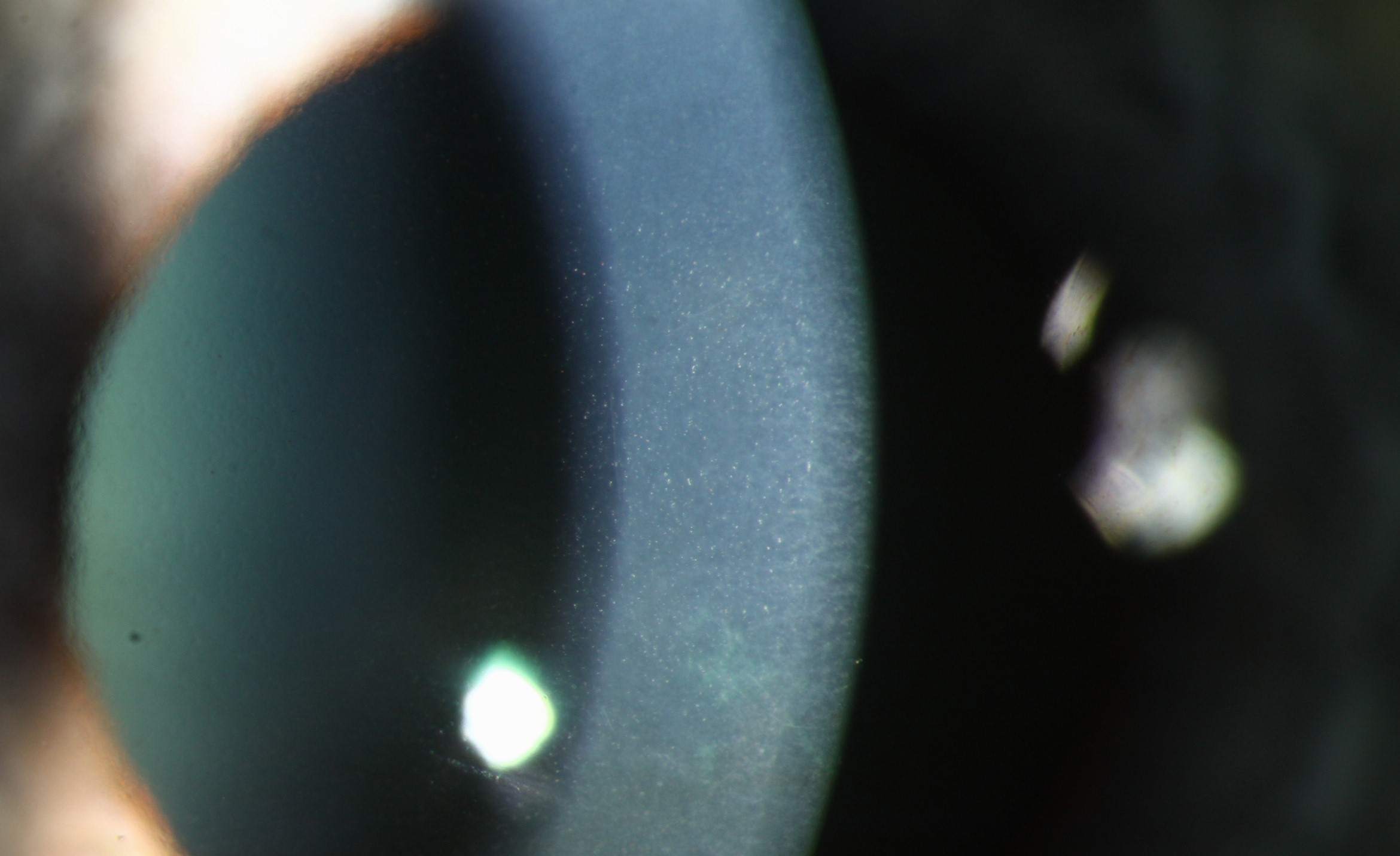
Corneal guttae represent focal excrescences of abnormal collagenous material deposited onto the posterior surface of Descemet membrane by dysfunctional endothelial cells, and their presence reflects an underlying, ongoing process of endothelial cell stress and gradual dropout, even when the remaining endothelial cell population is still functioning well enough to keep the cornea clear and edema-free.
The Beaten-Metal Appearance
With specular reflection technique at the slit lamp, a technique using a narrow, bright beam angled to catch the reflection off the corneal endothelial surface, guttae produce a characteristic dark, round, dropout pattern against the normally regular, mosaic-like endothelial cell reflection, often described as resembling hammered or beaten metal.
This specific examination technique, distinct from the standard diffuse or optic-section slit-lamp view most commonly used, is what makes early, sparse guttae visible, and clinicians should specifically employ specular reflection when endothelial assessment is a priority, since sparse early guttae can be easily missed with routine examination technique alone.
All-fit Slit-Lamp Adapter
Record and share exactly what you see at the slit lamp. One adapter fits any slit lamp or surgical microscope — and any smartphone.
From Choroida — the team behind this siteFuchs Endothelial Corneal Dystrophy
The presence of central corneal guttae, particularly with a family history and progression over time, is the earliest recognizable sign of Fuchs endothelial corneal dystrophy, and their detection, well before any corneal edema or visual symptoms develop, allows staging and monitoring of the condition from its earliest, asymptomatic phase (see Fuchs endothelial dystrophy).
Other Causes and Associations
- Age-related guttae, sometimes called Hassall-Henle bodies when located peripherally, represent a normal, common age-related finding of no clinical significance when confined to the far periphery, in contrast to the central guttae characteristic of Fuchs dystrophy
- Guttae can also develop secondary to chronic inflammation, prior intraocular surgery, or other causes of endothelial stress, distinct from the primary genetic dystrophy
Clinical Significance of Early Detection
- Guttae presence and density inform disease staging in Fuchs dystrophy, supporting more precise counseling about expected disease course
- Preoperative identification of guttae, particularly in a patient being evaluated for cataract surgery, is important since cataract surgery itself places additional stress on the endothelium, and eyes with a compromised endothelial reserve are at higher risk of postoperative corneal decompensation
- Specular microscopy, a more formal, quantitative technique for assessing endothelial cell density and morphology, is often used to further characterize endothelial health once guttae are identified on slit-lamp examination, providing objective numeric data to track over time
Monitoring
Patients with early, sparse guttae, particularly without any corneal edema or visual symptoms, are typically managed with periodic monitoring alone, since the disease may remain stable or progress only very slowly over many years, and premature intervention is not warranted based on the presence of guttae alone.
Progressive guttae density, development of stromal or epithelial edema, or emerging visual symptoms mark the transition to more advanced disease requiring active management (see corneal endothelial decompensation).
Preoperative Assessment Before Cataract Surgery
Given the additional endothelial stress that cataract surgery imposes, identifying guttae and assessing endothelial cell density preoperatively supports informed surgical planning and patient counseling, including discussion of the possibility that combined cataract and endothelial keratoplasty surgery, rather than cataract surgery alone, may ultimately be needed in eyes with significantly compromised endothelial reserve.
Prognosis
Early guttae detection, in itself, carries a generally favorable near-term prognosis, since many patients with sparse guttae never progress to visually significant disease within their lifetime.
The real value of early detection lies in the more informed monitoring and surgical planning it enables, allowing clinicians and patients to anticipate and plan for progressive endothelial disease, including surgical timing, well before symptoms force the issue.


Document what you see
Two smartphone imaging tools built for everyday clinic use — one for the slit lamp, one for the fundus.
From Choroida — the team behind this siteReferences
- Krachmer JH, Purcell JJ Jr, Young CW, Bucher KD. Corneal endothelial dystrophy. A study of 64 families. Arch Ophthalmol. 1978;96:2036-2039.
- Zoega GM, Fujisawa A, Sasaki H, et al. Prevalence and risk factors for cornea guttata in the Reykjavik Eye Study. Ophthalmology. 2006;113:565-569.
- Louttit MD, Kopplin LJ, Igo RP Jr, et al. A multicenter study to map genes for Fuchs endothelial corneal dystrophy: baseline characteristics and heritability. Cornea. 2012;31:26-35.
- Repp DJ, Hodge DO, Baratz KH, McLaren JW, Patel SV. Fuchs’ endothelial corneal dystrophy: subjective grading versus objective grading based on the central-to-peripheral thickness ratio. Ophthalmology. 2013;120:687-694.
