
Recurrent corneal erosion syndrome produces sudden, sharp eye pain on waking, night after night or in unpredictable episodes for months or years, as areas of corneal epithelium that never fully re-anchored after an initial injury or from an underlying dystrophy break down again with the mechanical stress of eyelid opening.
Treatment is genuinely stepwise, since most patients respond to simple measures and only a minority need a corneal procedure.
Why the Epithelium Keeps Breaking Down
The corneal epithelium normally anchors firmly to the underlying basement membrane and Bowman layer through hemidesmosomes and anchoring fibrils, a bond that takes weeks to months to fully mature after any injury that disrupts it.
If this anchoring complex fails to reform properly, whether from the original trauma itself, an underlying epithelial basement membrane dystrophy that produces an abnormal, poorly adherent basement membrane, or another anterior corneal dystrophy, the epithelium remains loosely attached, and the shearing force of the eyelid moving across the cornea during sleep, when the tear film is reduced and the eyelid can adhere to the epithelium, is often enough to pull it away.
Common Causes
- Prior corneal abrasion, particularly from a fingernail, paper edge, or plant material, which are classically associated with a higher risk of recurrent erosion than blunt or foreign body injuries
- Epithelial basement membrane dystrophy (map-dot-fingerprint dystrophy), the most common underlying dystrophy associated with spontaneous, non-traumatic recurrent erosion (see map-dot-fingerprint dystrophy)
- Other anterior corneal dystrophies, including some stromal dystrophies with anterior involvement
- Idiopathic cases, without an identified preceding trauma or dystrophy
All-fit Slit-Lamp Adapter
Record and share exactly what you see at the slit lamp. One adapter fits any slit lamp or surgical microscope — and any smartphone.
From Choroida — the team behind this siteClinical Presentation
- Sudden, sharp pain on waking or during the night, often with tearing, redness, and photophobia
- Episodes can range from mild discomfort resolving within minutes to severe pain persisting for a day or more with visible epithelial defect
- A history of a prior corneal abrasion, sometimes months to years earlier, or known epithelial basement membrane dystrophy
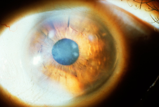
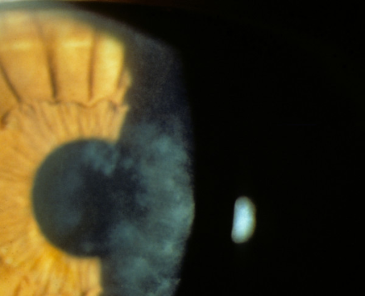
- Examination during an acute episode shows a focal epithelial defect or loose, heaped epithelium; between episodes, the cornea may look entirely normal or show subtle basement membrane changes on careful slit-lamp examination
Diagnosis
Diagnosis relies heavily on the characteristic history: recurrent, often nocturnal or early-morning pain, especially with a preceding relevant injury or known dystrophy.
Fluorescein staining during an acute episode confirms the epithelial defect, and careful examination between episodes for microcysts, fingerprint lines, or map-like patterns supports an underlying epithelial basement membrane dystrophy as the cause.
Stepwise Treatment
First-Line Medical Management
Most cases are managed successfully with medical therapy, aiming to support epithelial healing and reduce mechanical shear during sleep.
- Lubricating ointment at bedtime, to reduce eyelid-epithelium adherence overnight
- Hypertonic saline drops during the day and ointment at night, which can help reduce epithelial edema and improve adhesion
- A therapeutic bandage contact lens during an acute episode and sometimes for a period afterward, protecting the healing epithelium from eyelid shear
- Treatment of any acute defect with lubrication, topical antibiotic prophylaxis, and sometimes a bandage lens until the epithelium heals
Escalation for Refractory Cases
When erosions continue to recur despite an adequate trial of medical therapy, generally several months, procedural options are considered.
- Anterior stromal micropuncture, creating small, superficial puncture marks with a needle to promote firmer epithelial adhesion at the affected site, well suited to erosions outside the central visual axis given a theoretical small risk of subtle scarring
- Phototherapeutic keratectomy (PTK), using the excimer laser to smooth Bowman layer and promote stronger epithelial reattachment, effective for both traumatic and dystrophy-related recurrent erosion and suitable for central lesions as well
- Diamond burr polishing of Bowman layer, a mechanical alternative to laser PTK with similar goals
Choosing Among Procedures
The choice depends on the location and extent of the erosion-prone area, the presence of an underlying dystrophy affecting a broader area of cornea, and surgeon and patient preference; PTK is generally favored when an underlying dystrophy affects a larger corneal area, since it can treat a wider zone more uniformly than micropuncture.
Prognosis
The majority of patients respond well to medical therapy alone, and most who require a procedure achieve durable resolution of their erosion episodes afterward.
Recurrence after procedural treatment is possible but uncommon, and patients with widespread epithelial basement membrane dystrophy may need broader or repeat treatment if new areas become symptomatic over time.


Document what you see
Two smartphone imaging tools built for everyday clinic use — one for the slit lamp, one for the fundus.
From Choroida — the team behind this siteReferences
- Ramamurthi S, Rahman MQ, Dutton GN, Ramaesh K. Pathogenesis, clinical features and management of recurrent corneal erosions. Eye (Lond). 2006;20:635-644.
- Das S, Seitz B. Recurrent corneal erosion syndrome. Surv Ophthalmol. 2008;53:3-15.
- Dua HS, Gomes JA, Singh A. Corneal epithelial wound healing. Br J Ophthalmol. 1994;78:401-408.
- Soong HK, Farjo AA, Katz D, Meyer RF, Sugar A. Diamond burr superficial keratectomy for recurrent corneal erosions. Br J Ophthalmol. 2002;86:296-298.
