
Pneumatic retinopexy repairs select rhegmatogenous retinal detachments by injecting a gas bubble into the vitreous cavity to tamponade a retinal break from the inside, combined with laser or cryotherapy to seal the break, all performed in an office or minor procedure setting rather than a full operating room.
It offers a genuinely different risk-benefit trade-off from scleral buckle surgery or pars plana vitrectomy, both discussed in their own dedicated articles on this site: a much less invasive procedure, but one that only works reliably for a specific, favorable subset of detachments.
Understanding exactly which detachments qualify for this approach, and why patient positioning after the procedure matters so much, is central to using pneumatic retinopexy successfully rather than as a poor substitute for a more invasive repair.
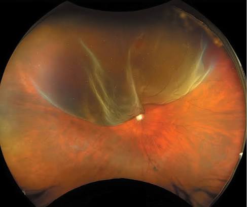
Mechanism
A gas bubble injected into the vitreous cavity rises to tamponade a superior retinal break from the inside, sealing it against the underlying retinal pigment epithelium and preventing further fluid from tracking under the retina while the laser or cryotherapy treatment forms a permanent chorioretinal adhesion around the break.
Because the bubble must rise to cover the specific break causing the detachment, precise patient positioning after the injection is essential, and the technique depends on breaks located in the superior retina where a rising bubble can actually reach and tamponade them.
Once the chorioretinal adhesion has matured, generally over several days, the bubble’s job is essentially done, and it gradually resorbs on its own over the following one to several weeks depending on which gas was used.
Patient Selection
- A single break, or a small cluster of breaks confined to a limited area, located in the superior two-thirds of the retina where a rising gas bubble can reliably tamponade it
- Clear ocular media, allowing adequate visualization to confirm all breaks have been identified before the procedure, since a missed break elsewhere in the retina will not be addressed by a bubble positioned over a different area
- A patient able to understand and reliably maintain the specific positioning regimen required after the procedure, since positioning compliance directly determines whether the bubble tamponades the correct area
- Absence of significant proliferative vitreoretinopathy or other complicating factors that would favor a more comprehensive surgical approach from the outset
Inferior breaks, multiple breaks spread across different quadrants, and significant proliferative vitreoretinopathy generally make pneumatic retinopexy a poor choice, favoring scleral buckle or vitrectomy instead.
Fundus Explorer Pro
Photograph the retinal findings described here with the phone already in your pocket — 22 D optics and built-in illumination in one handheld unit.
From Choroida — the team behind this siteProcedure and Positioning
The procedure is typically performed at the slit lamp or in a minor procedure room, with the gas bubble injected through the pars plana after appropriate anesthesia and often after a small amount of aqueous is removed to accommodate the injected gas volume.
Laser retinopexy or cryotherapy, both discussed in their own dedicated articles on this site, is applied around the break either just before or shortly after the gas injection, depending on the specific protocol and view available at the time.
Strict positioning instructions follow, requiring the patient to hold their head in a specific orientation, often for much of the day over several days, to keep the bubble tamponading the correct area of retina while the chorioretinal adhesion matures.
Advantages and Limitations
The office-based, minimally invasive nature of the procedure is its central appeal, avoiding an operating room, general anesthesia, and the longer recovery associated with scleral buckle or vitrectomy.
Success rates with a single procedure are generally somewhat lower than with scleral buckle or vitrectomy, particularly when patient selection is imperfect or positioning compliance is inconsistent, and a failed pneumatic retinopexy typically still requires a subsequent, more definitive surgical repair.
Cataract progression, a concern with vitrectomy, discussed in its own dedicated article on this site, is largely avoided with pneumatic retinopexy, which is a meaningful consideration in phakic patients weighing their options.
Complications
New or missed retinal breaks can occur or become apparent during the postoperative period, sometimes related to the same vitreous traction that caused the original detachment, requiring prompt additional treatment if identified.
Subretinal gas migration, if the gas inadvertently passes beneath the retina rather than remaining in the vitreous cavity, is a recognized complication that compromises the intended tamponade effect.
Elevated intraocular pressure from the injected gas volume is common in the immediate postprocedure period and is monitored and managed accordingly.


Document what you see
Two smartphone imaging tools built for everyday clinic use — one for the slit lamp, one for the fundus.
From Choroida — the team behind this siteReferences
- Hilton GF, Grizzard WS. Pneumatic retinopexy: a two-step outpatient operation without conjunctival incision. Ophthalmology.
- American Academy of Ophthalmology. Basic and Clinical Science Course, Section 12: Retina and Vitreous.
- Hillier RJ, Felfeli T, Berger AR, et al. The Pneumatic Retinopexy versus Vitrectomy for the Management of Primary Rhegmatogenous Retinal Detachment Outcomes Randomized Trial (PIVOT). Ophthalmology.
