
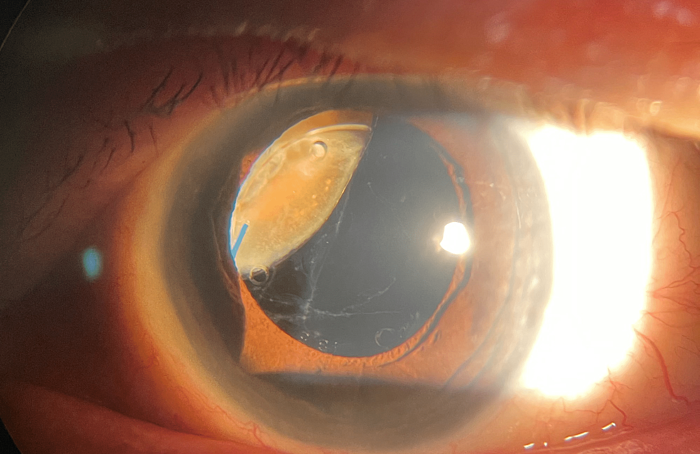
Retained lens fragments, commonly called a dropped nucleus, occur when a piece of the natural lens falls posteriorly into the vitreous cavity during complicated cataract surgery, most often following unexpected posterior capsule rupture.
It represents a genuinely different clinical scenario from lens-particle glaucoma, discussed in its own dedicated article on this site, which addresses smaller cortical fragments causing outflow obstruction in the anterior chamber: a dropped nucleus is a larger fragment that has migrated all the way into the vitreous cavity itself, well behind the iris and out of the anterior segment surgeon’s normal reach.
Recognizing this distinction, and understanding why a dropped nucleus almost always requires a posterior segment surgeon rather than being managed by the original anterior segment surgeon alone, is central to handling this complication appropriately.
Mechanism
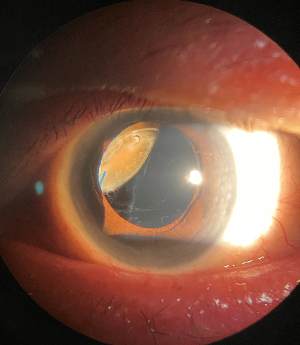
Posterior capsule rupture during phacoemulsification, discussed in its own dedicated article on this site, creates an opening through which lens material can fall posteriorly rather than remaining accessible in the anterior segment where the surgery was being performed.
Once nuclear or cortical material passes behind an intact or partially intact vitreous face, blind attempts to retrieve it with anterior segment instruments risk further vitreous traction, additional capsular damage, and a meaningfully increased risk of retinal complications from that traction.
This is precisely why the standard, safest approach shifts at this point from continued attempts at anterior retrieval to referral for a formal pars plana vitrectomy, discussed in its own dedicated article on this site, performed by a surgeon experienced in posterior segment technique.
Clinical Significance
Retained lens material is not inert once inside the eye: it provokes inflammation, and larger or denser retained fragments in particular can cause significant intraocular inflammation and elevated pressure if left untreated for an extended period.
The inflammatory response to retained lens material can, over time, contribute to cystoid macular edema, glaucoma, and chronic uveitis, adding to the visual risk beyond whatever complications occurred during the index surgery itself.
Small, soft cortical remnants are sometimes managed expectantly with close observation, since minimal fragments occasionally clear with medical anti-inflammatory management alone, but denser nuclear fragments generally do not resolve on their own and require surgical removal.
Fundus Explorer Pro
Photograph the retinal findings described here with the phone already in your pocket — 22 D optics and built-in illumination in one handheld unit.
From Choroida — the team behind this siteManagement
- Prompt referral to a vitreoretinal surgeon once a dropped nucleus is recognized, rather than prolonged attempts at anterior segment retrieval that risk worsening the situation
- Topical corticosteroids and aqueous suppressants in the interim, to control inflammation and pressure while awaiting definitive surgical management
- Pars plana vitrectomy to retrieve and remove the retained lens material, typically performed within one to two weeks of the original surgery once the eye has had some time to stabilize, though timing is individualized based on the clinical picture
- Placement or exchange of an intraocular lens, either at the time of the vitrectomy or as a separate, later procedure, depending on the specific circumstances of the case
Timing Considerations
Very early intervention, within the first day or two, is sometimes favored for large, dense fragments causing significant acute inflammation or pressure elevation that is not adequately controlled medically.
A short delay of one to two weeks is more typical for less urgent cases, allowing initial postoperative inflammation from the index surgery to settle somewhat before undertaking a second procedure, which can make the vitrectomy technically more straightforward.
Excessive delay, on the other hand, risks prolonged inflammation and its downstream complications, so the interval is a deliberate balance rather than simply “as soon as possible” or “whenever convenient.”
Outcomes
Visual outcomes after appropriately managed retained lens fragment removal are often good, particularly when the retained material is identified and referred promptly and the vitrectomy is performed without further complication.
Outcomes are less favorable when there has been a significant delay in recognition or referral, when the retained fragment burden was large, or when the original complicated surgery involved additional complications such as significant vitreous loss or zonular compromise beyond the capsule rupture alone.
Clear, honest communication with the patient about what happened, why a second procedure and a different surgeon are needed, and what the revised visual prognosis realistically looks like is an important part of managing this complication well, beyond the surgical technique itself.


Document what you see
Two smartphone imaging tools built for everyday clinic use — one for the slit lamp, one for the fundus.
From Choroida — the team behind this siteReferences
- Vilar NF, Flynn HW Jr, Smiddy WE, et al. Removal of retained lens fragments after phacoemulsification reverses secondary glaucoma and restores visual acuity. Ophthalmology.
- American Academy of Ophthalmology. Basic and Clinical Science Course, Section 11: Lens and Cataract.
- Ho LY, Doft BH, Wang L, Bunker CH. Clinical predictors and outcomes of pars plana vitrectomy for retained lens material after cataract extraction. American Journal of Ophthalmology.
