
Amniotic membrane transplantation uses processed placental tissue, rich in growth factors and possessing natural anti-inflammatory and anti-scarring properties, to promote healing of a damaged or diseased ocular surface.
It has become one of the more versatile tools available for ocular surface reconstruction, used across a wide range of conditions from acute chemical burns to chronic non-healing epithelial defects, precisely because its biological properties support healing rather than simply providing a mechanical barrier or patch.
Understanding what the membrane actually does biologically, rather than thinking of it as an inert covering, explains why it has found such broad application across genuinely different categories of ocular surface disease.
Donor tissue is screened and processed under strict protocols before clinical use, and both fresh and preserved (typically cryopreserved or dehydrated) forms are available, with the choice between them driven by institutional availability and specific clinical preference.
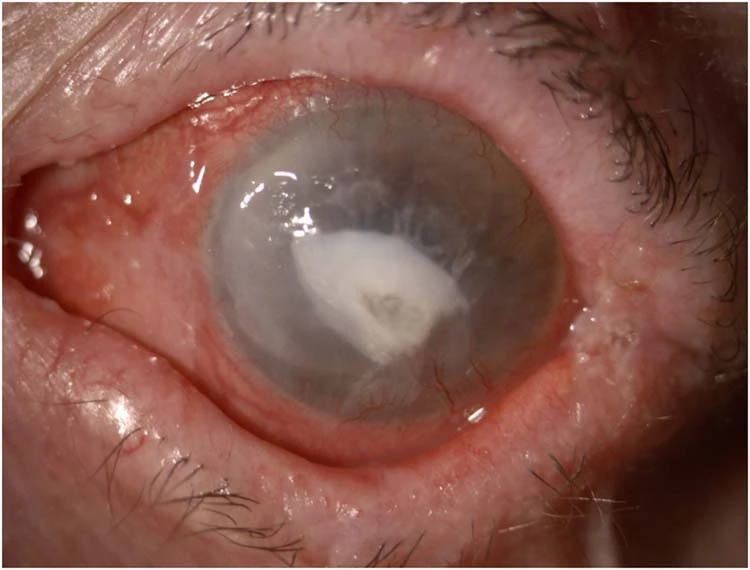
What the Membrane Does
Amniotic membrane, harvested from the innermost layer of the placenta after an elective cesarean delivery and processed for clinical use, contains a basement membrane and an avascular stromal matrix rich in growth factors, cytokines, and anti-inflammatory and anti-fibrotic proteins.
Placed against a damaged ocular surface, the membrane supports epithelial cell migration and adhesion, suppresses excessive inflammation, and reduces the scarring and fibrosis that would otherwise complicate healing of a significant surface injury.
This combination of properties, essentially providing a biologically favorable scaffold for the eye’s own healing rather than an inert covering, is what distinguishes it from a simple bandage contact lens or patch.
Indications
- Acute chemical or thermal burns, discussed in relation to chemical eye injury in its own dedicated article on this site, where early amniotic membrane placement can reduce scarring and limbal stem cell loss
- Persistent epithelial defects that have failed to heal with conventional measures such as lubrication and bandage contact lens use
- Neurotrophic keratopathy, discussed in its own dedicated article on this site, where impaired healing from reduced corneal sensation makes the membrane’s pro-healing properties particularly useful
- Pterygium surgery, as an alternative or adjunct to conjunctival autografting, discussed in its own dedicated article on this site, in select cases
- Symblepharon prevention or treatment after severe conjunctival injury or surgery, helping maintain normal fornix architecture during healing
All-fit Slit-Lamp Adapter
Record and share exactly what you see at the slit lamp. One adapter fits any slit lamp or surgical microscope — and any smartphone.
From Choroida — the team behind this siteApplication Techniques
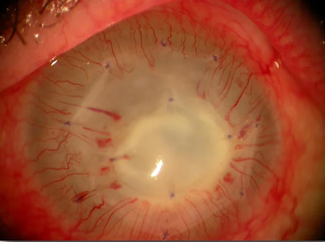
The membrane can be applied as a graft, sutured or glued directly onto a defect to promote healing at that specific site, or as a patch, placed over the entire ocular surface to provide broader anti-inflammatory and protective coverage.
A sutureless option using a symblepharon ring to hold the membrane in place against the ocular surface offers a faster, office-based application for appropriate cases, avoiding the need for suturing in a more urgent or less invasive setting.
The choice between graft and patch technique, and between sutured and sutureless application, depends on the specific indication, the size and depth of the area needing treatment, and practical considerations including the clinical setting and urgency.
Placement orientation, with the basement membrane side facing up or down depending on the goal of the treatment, is a technical detail surgeons attend to carefully, since it affects how the tissue interacts with the underlying ocular surface.
In Acute Chemical Injury Specifically
Early amniotic membrane application after a significant chemical burn can meaningfully reduce the inflammatory cascade and subsequent scarring that would otherwise threaten both corneal clarity and limbal stem cell survival.
Timing matters considerably in this setting, since the membrane’s anti-inflammatory benefit is most valuable when applied early in the acute injury course, before extensive scarring and stem cell loss have already occurred.
This makes amniotic membrane one of the more time-sensitive interventions available for severe chemical injury, alongside the emergency irrigation and other acute measures covered in the dedicated chemical eye injury article on this site.
Outcomes and Limitations
Amniotic membrane transplantation promotes healing and reduces scarring effectively across many of its indications, but it does not replace the need to address an underlying cause, such as ongoing exposure, infection, or active autoimmune disease, which must still be managed directly.
The membrane itself is gradually resorbed or sloughed as the underlying epithelium heals, meaning its effect is time-limited by design, supporting the healing process rather than providing a permanent structural replacement.
For severe or extensive limbal stem cell loss, amniotic membrane alone is generally insufficient, and more substantial interventions, including limbal stem cell transplantation, are needed to restore a functional ocular surface.
Setting realistic expectations before use matters here, since patients and even some clinicians occasionally overestimate what the membrane alone can accomplish in a severely compromised ocular surface.


Document what you see
Two smartphone imaging tools built for everyday clinic use — one for the slit lamp, one for the fundus.
From Choroida — the team behind this siteReferences
- Dua HS, Gomes JA, King AJ, Maharajan VS. The amniotic membrane in ophthalmology. Survey of Ophthalmology.
- American Academy of Ophthalmology. Basic and Clinical Science Course, Section 8: External Disease and Cornea.
- Kheirkhah A, Casas V, Raju VK, Tseng SC. Sutureless amniotic membrane transplantation for partial limbal stem cell deficiency. American Journal of Ophthalmology.
