
CASE REPORT
A 65-year-old female visited an eye clinic with a presenting complaint of decreased vision in her right eye. On testing Visual acuity, it was recorded as OD: 6/36, OS: 6/9. On a fundus examination, raised mulberry-like lesions were seen.
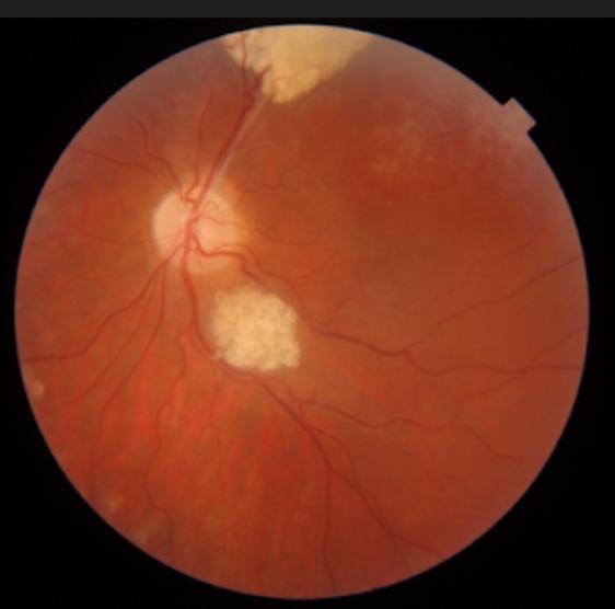
On OCT, subretinal fluid was also noticeable. This patient was treated with bevacizumab as the first treatment of choice and it resulted in significant improvement in her visual acuity. Genetic testing was done to rule out its leading Aetiology of either tuberous sclerosis complex, neurofibromatosis, or others but it was found negative.
Based on fundus findings and OCT report, the diagnosis was confirmed as a retinal astrocytic hamartoma.
Retinal astrocytic hamartoma entity
Retinal astrocytic hamartoma is a benign retinal tumor composed of astrocytic glial cells having frequent association with tuberous sclerosis complex(TSC) and rare association with neurofibromatosis.
Other associations are as follows:
- Retinitis pigmentosa (RP)
- Stargardt’s disease
- Gyrate atrophy
- Usher’s syndrome
In 1921, it was recognized first time by Van der Hoeve who named the lesions(tumors) “phakomas”.
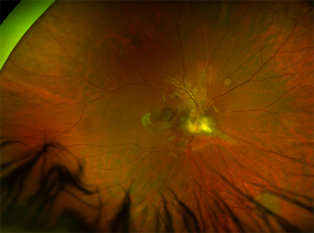
Retinal astrocytic hamartoma patients are usually asymptomatic but if lesions(tumors) are present in the macular or optic nerve region, it can lead to decreased vision, Metamorphopsia, or floaters. Fundus findings range from flat, smooth grey-white lesions to raised, multilobulated, calcified white-yellow lesions.
Morphologically, retinal astrocytic hamartoma can be classified into 3 types.
Type 1:
In this type, non-calcified grey-white lesions are seen which are flat and smooth. It is the most commonly occurring type.
Type 2:
In this type, mulberry-like lesions are seen which are multinodular, raised, and calcified as well.
Type 3:
This type of retinal astrocytic hamartoma has mixed signs of both type 1 and type 2.
Fundus Explorer Pro
Photograph the retinal findings described here with the phone already in your pocket — 22 D optics and built-in illumination in one handheld unit.
From Choroida — the team behind this siteDiagnostic procedures
It can be misdiagnosed with retinoblastoma so a B scan and OCT help distinguish retinal astrocytic hamartoma from retinoblastoma
OCT:
Spectral-domain (SD) OCT is thought to be more effective in diagnosing RAH. It shows dome-shaped thickening of the inner retina and intralesional calcification in the form of empty spaces. The outer retina might be compressed or disorganized by the tumor.
Ultrasound:
It demonstrates the tumor as well as hyperechogenic calcifications.
Fundus Autofluorescence(FAF):
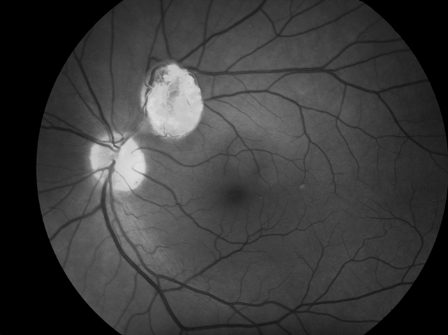
It is done to differentiate between calcified and non-calcified tumors. Calcified tumors show hyper autofluorescence and non-calcified tumors show reduced autofluorescence.
Fluorescein Angiography:
It helps identify those tumors which are not visible clinically.
MANAGEMENT
Most retinal astrocytic hamartomas are asymptomatic and need only observation; anti-VEGF agents such as bevacizumab are used for lesions that affect vision. mTOR Inhibitors (sirolimus or everolimus) are usually used in patients with TSC.
In the case of aggressive RAH, other therapies can be used such as photodynamic therapy, laser photocoagulation, and Dexamethasone implant.



Document what you see
Two smartphone imaging tools built for everyday clinic use — one for the slit lamp, one for the fundus.
From Choroida — the team behind this siteREFERENCES
- Amoroso F, Souied EH, Bruyère E, Astroz P, Mouallem-Beziere A, Pedinielli A, Miere A. Two-year follow-up of a retinal astrocytic hamartoma imaged by optical coherence tomography angiography. Retin Cases Brief Rep. 2020 Jan 29; doi: 10.1097/ICB.0000000000000975.
- Bennett LW. Isolated retinal astrocytic hamartoma. Clin Exp Optom. 2020 May;103(3):382–383. doi: 10.1111/cxo.12956.
- Zhang P, Sun D, Zhu J, Li J, Wang Y. Image features of retinal astrocytic hamartoma in a patient with tuberous sclerosis complex. Eye Sci. 2014 Dec;29(4):223–226.
- Shields JA, Shields CL. Glial tumors of the retina and optic disc. In: Shields JA, Shields CL, editors. Intraocular tumors. A Text and Atlas. Philadelphia: WB Saunders; 2008. pp. 405–27.
- Nyboer JH, Robertson DM, Gomer MR. Retinal lesions in tuberous sclerosis. Arch Ophthalmol. 1976;94:1277–80.
- Crino P, Nathanson K, Henske E. The tuberous sclerosis complex. N Engl J Med. 2006;355:1345–56.
Test yourself
A few questions straight from this article.
-
What type of cell makes up a retinal astrocytic hamartoma?
Retinal astrocytic hamartoma is a benign retinal tumour composed of astrocytic glial cells. -
Which systemic disorder is most frequently associated with retinal astrocytic hamartoma?
The tumour has a frequent association with tuberous sclerosis complex, while its association with neurofibromatosis is rare. -
When does a retinal astrocytic hamartoma cause decreased vision, metamorphopsia or floaters?
Most patients are asymptomatic; symptoms such as decreased vision, metamorphopsia or floaters appear when lesions occupy the macular or optic nerve region. -
What does the most common morphological type of retinal astrocytic hamartoma look like?
Type 1, the most common type, is a flat, smooth, non-calcified grey-white lesion; type 2 is the raised calcified mulberry lesion and type 3 mixes both. -
What does fluorescein angiography contribute in retinal astrocytic hamartoma?
Fluorescein angiography helps identify tumours that cannot be seen clinically; separating calcified from non-calcified tumours is the role of autofluorescence. -
Retinal astrocytic hamartoma can be misdiagnosed as which tumour, making B-scan and OCT useful?
Retinal astrocytic hamartoma can be mistaken for retinoblastoma, so B-scan ultrasound and OCT are used to tell the two apart. -
What does spectral-domain OCT typically show in retinal astrocytic hamartoma?
SD-OCT shows dome-shaped thickening of the inner retina with intralesional calcification appearing as empty spaces, and the outer retina may be compressed or disorganised. -
How do calcified retinal astrocytic hamartomas appear on fundus autofluorescence?
Autofluorescence separates the two forms: calcified tumours are hyperautofluorescent, whereas non-calcified tumours show reduced autofluorescence. -
Which drug class is usually used for retinal astrocytic hamartoma in patients with tuberous sclerosis complex?
mTOR inhibitors such as sirolimus or everolimus are usually used in patients who have tuberous sclerosis complex. -
Which therapies are listed for aggressive retinal astrocytic hamartoma?
For aggressive lesions the article lists photodynamic therapy, laser photocoagulation and a dexamethasone implant as further options.
