
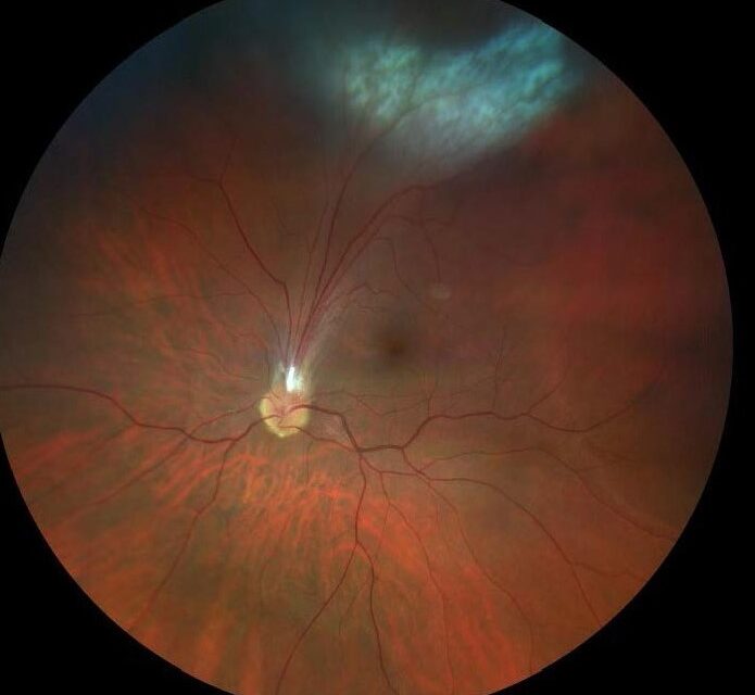
Ocular toxocariasis is intraocular inflammation and granuloma formation caused by larval migration of Toxocara species, most commonly Toxocara canis, an intestinal roundworm of dogs that can infect humans as an accidental, dead-end host.
It is a classic pediatric uveitis mimic and one of the important entities to consider whenever a child presents with unilateral leukocoria, vision loss, or a retinal mass, since it can closely resemble far more ominous diagnoses on initial presentation.
Distinguishing ocular toxocariasis from retinoblastoma in particular is one of the more consequential differential diagnoses in pediatric ophthalmology, given how different the management and prognosis are for the two conditions.
Unlike ocular toxoplasmosis, discussed in its own dedicated article on this site, ocular toxocariasis is caused by a different organism entirely, an intestinal nematode rather than a protozoan, and the two should not be confused despite the similarity of their names.
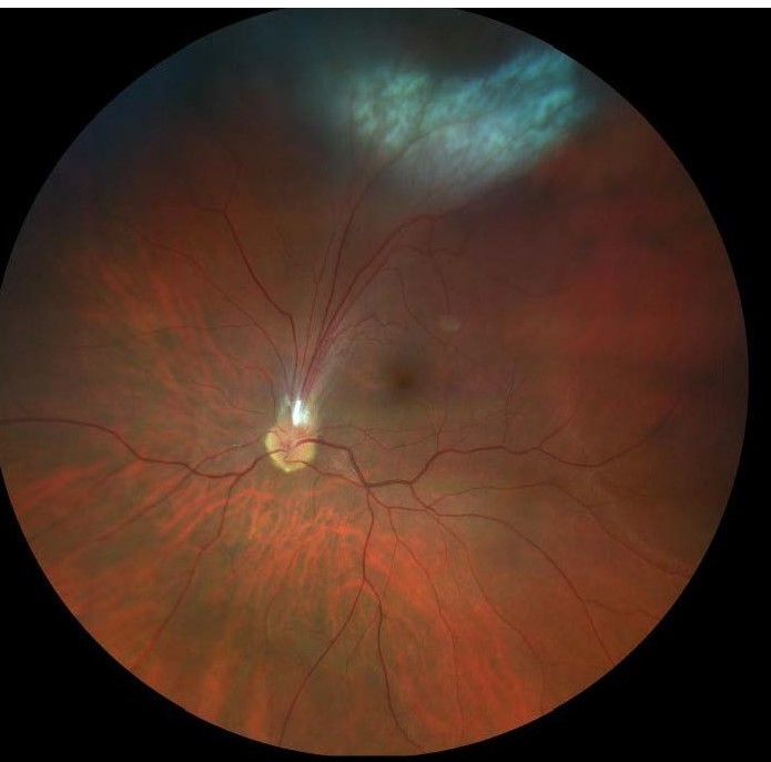
Transmission and Pathophysiology
Infection follows ingestion of Toxocara eggs from soil, sand, or contaminated surfaces contaminated by dog or cat feces, most commonly in young children with pica or frequent hand-to-mouth contact during outdoor play.
Ingested eggs hatch into larvae in the intestine, which then migrate through the bloodstream to various tissues, including, in a minority of infected individuals, the eye, where they die and provoke a granulomatous inflammatory response rather than completing their normal life cycle as they would in a dog.
Ocular involvement typically occurs without the more classic systemic visceral larva migrans picture of hepatomegaly, pulmonary symptoms, and eosinophilia, which is part of why ocular toxocariasis is often an isolated finding without an obvious preceding systemic illness to point toward the diagnosis.
The typical age at presentation is between five and ten years, though it can occur outside this range, and a history of close contact with young, unwormed puppies is a common but not universal feature.
Clinical Presentation
- Unilateral vision loss or strabismus in a child, often the presenting complaint that brings the family to an eye exam
- A peripheral granuloma, a localized, elevated white or yellow-white lesion in the peripheral retina, sometimes with an associated fibrous band extending toward the optic disc
- A posterior pole granuloma, involving the macula directly and causing more significant central vision loss than the peripheral form
- Diffuse chronic endophthalmitis, a more severe presentation with vitritis, retinal detachment, and a leukocoric appearance that can closely mimic retinoblastoma
Fundus Explorer Pro
Photograph the retinal findings described here with the phone already in your pocket — 22 D optics and built-in illumination in one handheld unit.
From Choroida — the team behind this siteWhy It Is a Retinoblastoma Mimic
Both conditions can present in a young child with leukocoria, a white pupillary reflex, and both can produce a mass-like lesion on fundus exam, which is exactly why ocular toxocariasis sits prominently on the differential for any child presenting this way.
B-scan ultrasonography, discussed in its own dedicated article on this site, is useful for distinguishing the two: retinoblastoma classically shows calcification on ultrasound, a feature not typical of a toxocara granuloma.
Serologic testing for Toxocara antibodies, along with a careful history for pica, geophagia, or pet exposure, supports the diagnosis, though a definitive distinction from retinoblastoma sometimes requires the full range of ancillary testing available in a specialized ocular oncology setting given the stakes of missing a malignancy.
Diagnostic Evaluation
The diagnosis is often made from the characteristic clinical appearance combined with supportive serology, though serologic testing can be falsely negative, particularly when the infectious burden was low or the exposure was remote in time.
Imaging, including B-scan ultrasonography and, when available, optical coherence tomography, helps characterize the granuloma and any associated traction or fibrous proliferation extending across the vitreous.
A thorough exclusion of retinoblastoma is essential in any ambiguous case, given the fundamentally different stakes and management pathways for the two conditions.
Management
Systemic or periocular corticosteroids are used to control active inflammation, particularly in the diffuse endophthalmitis form, aiming to limit ongoing tissue damage from the inflammatory response to the dying larva.
Antihelminthic medications have a more limited and debated role in established ocular disease, since the inflammatory damage is largely driven by the immune response to a larva that has typically already died, rather than by an ongoing active infection needing eradication.
Surgical intervention, including vitrectomy, may be needed for complications such as tractional retinal detachment or dense vitreous membranes resulting from chronic inflammation and fibrosis.
Prevention through routine deworming of pet dogs and cats, along with basic hygiene measures in young children, remains the most effective public health approach to reducing the overall burden of this preventable condition.
Long-term follow-up matters even after inflammation quiets down, since a stable granuloma can still contract fibrous tissue over subsequent years, occasionally producing a late tractional retinal detachment well after the initial presentation.


Document what you see
Two smartphone imaging tools built for everyday clinic use — one for the slit lamp, one for the fundus.
From Choroida — the team behind this siteReferences
- Shields JA, Shields CL. Ocular toxocariasis: a review. Survey of Ophthalmology.
- American Academy of Ophthalmology. Basic and Clinical Science Course, Section 9: Uveitis and Ocular Inflammation.
- Ahn SJ, Woo SJ, Jin Y, et al. Clinical features and course of ocular toxocariasis in adults. PLOS Neglected Tropical Diseases.
