
Traumatic optic neuropathy is optic nerve injury following blunt or penetrating trauma to the head, face, or orbit, and it is one of the few causes of vision loss where the fundus can look completely normal at the moment the damage is occurring.
The nerve is most often injured indirectly, from a force transmitted to it rather than a direct laceration, which is part of why the diagnosis is so easy to miss in a trauma bay focused on more obviously life-threatening injuries.
A normal-appearing disc with markedly reduced vision after facial trauma should always raise this diagnosis rather than be dismissed as a lesser injury.
It occurs in a small but meaningful proportion of patients with significant head trauma, and it is frequently overshadowed by more visible or more immediately life-threatening injuries in the same patient.
Mechanism
Indirect traumatic optic neuropathy typically results from a deceleration force transmitted through the bony orbit to the optic canal, where the nerve is fixed and relatively unable to move, making it vulnerable to shearing and contusion injury even without any fracture being visible.
Direct injury is less common and results from actual penetration, laceration, or bony impingement on the nerve itself, often from a displaced fracture fragment or a penetrating foreign body.
Secondary injury from edema and ischemia within the tight confines of the optic canal can compound the initial mechanical insult, which is part of the rationale behind decompression as a theoretical treatment option, discussed further below.
Clinical Presentation
- Sudden vision loss after head or facial trauma, ranging from mild to complete, occurring immediately or, less commonly, in a delayed fashion over hours to days
- A relative afferent pupillary defect on the affected side, present even when the fundus looks entirely normal, since the injury is retrobulbar
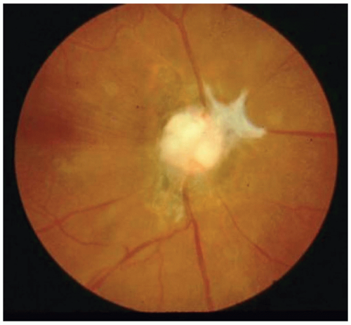
- A normal-appearing optic disc acutely, with pallor developing only weeks later if the axonal injury is severe enough to cause degeneration
- Associated signs of orbital or craniofacial trauma, including periorbital ecchymosis, orbital fracture, or reduced consciousness that can make the ophthalmic exam more difficult to perform reliably
The combination of profound vision loss with a completely unremarkable fundus exam is the classic diagnostic clue, and it is precisely what makes this injury easy to overlook without deliberate testing of the pupillary light reflex.
Fundus Explorer Pro
Photograph the retinal findings described here with the phone already in your pocket — 22 D optics and built-in illumination in one handheld unit.
From Choroida — the team behind this siteEvaluation
A relative afferent pupillary defect must be checked specifically, since it is often the only objective sign of the injury in the first hours after trauma, well before any disc changes appear.
Thin-section CT of the orbit and optic canal is the standard imaging study, looking for a canal fracture, bony impingement on the nerve, or a retrobulbar hematoma that might be amenable to surgical decompression.
Formal visual field testing and color vision assessment, when the patient’s condition and cooperation allow, add useful baseline documentation for tracking any change over time.
Visual evoked potentials can offer objective evidence of nerve function in a patient too obtunded to cooperate with subjective testing, though they are used selectively rather than as a routine part of every workup.
Management
High-dose corticosteroids were used for years based on extrapolation from spinal cord injury data, but the International Optic Nerve Trauma Study and subsequent literature failed to show a clear visual benefit, and steroids at those doses carry real systemic risk in an already traumatized patient.
Current practice has shifted away from routine high-dose steroids for this indication, with treatment decisions made on a case-by-case basis rather than as a default protocol.
Surgical optic canal decompression is reserved for selected cases, generally when there is clear bony compression on imaging and either a delayed or progressive vision loss pattern, since the evidence for decompression improving outcomes in a stable, complete injury from the outset is weak.
Most cases are managed with observation and close monitoring for progression, coordinating with the trauma and neurosurgical teams managing the patient’s other injuries.
Prognosis
Visual outcome correlates most strongly with the severity of vision loss at presentation, and a patient who presents with no light perception has a substantially lower chance of meaningful recovery than one who retains some vision initially.
Some spontaneous improvement can occur over the following weeks even without specific treatment, which is one reason outcomes from case series with variable treatment protocols have been difficult to interpret cleanly.
Family counseling should be honest about this uncertainty from the outset, since the range of plausible outcomes for a given patient is genuinely wide and depends on factors that are only partly measurable at the time of initial evaluation.
Long-term follow-up eventually shows the disc pallor that acute imaging and exam findings could not predict early on, and that pallor becomes the permanent structural marker of the original nerve injury.


Document what you see
Two smartphone imaging tools built for everyday clinic use — one for the slit lamp, one for the fundus.
From Choroida — the team behind this siteReferences
- Levin LA, Beck RW, Joseph MP, et al. The treatment of traumatic optic neuropathy: the International Optic Nerve Trauma Study. Ophthalmology.
- Steinsapir KD, Goldberg RA. Traumatic optic neuropathy: an evolving understanding. American Journal of Ophthalmology.
- American Academy of Ophthalmology. Basic and Clinical Science Course, Section 5: Neuro-Ophthalmology.
- Yu-Wai-Man P, Griffiths PG. Steroids for traumatic optic neuropathy. Cochrane Database of Systematic Reviews.
