
Case Study
A 38-year-old male presented with mild blurred vision in the left eye. The patient denied any pain, photophobia, or systemic symptoms. Visual acuity was 20/20 in both eyes.
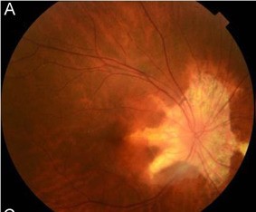
Fundus examination revealed peripapillary atrophic lesions extending in a radial, helicoid pattern from the optic disc toward the mid-periphery in the left eye.
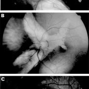
The right eye was unremarkable. Optical coherence tomography (OCT) demonstrated areas of retinal pigment epithelium (RPE) and choriocapillaris atrophy along the helicoid lesions.
Fluorescein angiography confirmed window defects corresponding to the atrophic zones. There was no evidence of inflammation or active leakage.
A diagnosis of unilateral helicoid peripapillary chorioretinal degeneration (HPCD) was made.
Disease Entity
Helicoid Peripapillary Chorioretinal Degeneration (HPCD) is a rare, non-inflammatory degenerative retinal condition characterized by curvilinear, helicoid zones of chorioretinal atrophy radiating from the optic disc.
It most often presents asymptomatically and is discovered incidentally during routine ophthalmologic evaluation.
Fundus Explorer Pro
Photograph the retinal findings described here with the phone already in your pocket — 22 D optics and built-in illumination in one handheld unit.
From Choroida — the team behind this siteEpidemiology
HPCD is extremely rare and has been reported sporadically in the literature. It is more commonly seen in middle-aged individuals and may be unilateral or bilateral.
There is no known gender or racial predilection. Familial cases are exceedingly rare.
Pathophysiology
The exact pathogenesis of HPCD remains unclear. It is presumed to involve chronic, progressive atrophy of the RPE and choriocapillaris along specific retinal nerve fiber layer pathways, possibly related to congenital or developmental defects.
Unlike inflammatory chorioretinal disorders, HPCD lacks signs of active inflammation or immune-mediated damage.
Clinical Features
-
Symptoms: Often asymptomatic
-
Visual Acuity: Typically preserved unless the macula is involved
-
Fundus Findings:
-
Peripapillary helicoid zones of chorioretinal atrophy
-
Retinal pigment mottling
-
Clear demarcation between healthy and atrophic retina
-
Absence of hemorrhage or exudation
-
Examination Findings
-
OCT: Thinning and disruption of outer retinal layers and RPE in helicoid patterns
-
Fundus Autofluorescence: Hypoautofluorescent zones corresponding to atrophy
-
Fluorescein Angiography: Window defects due to RPE loss
-
Visual Fields: May show localized defects correlating with atrophic tracks
Differential Diagnosis
-
Peripapillary atrophy from myopia
-
Retinitis pigmentosa (especially pericentral type)
-
Toxoplasmosis scars
-
Chorioretinal scars from previous inflammation
-
Serpiginous choroiditis (although HPCD lacks inflammation)
Diagnosis
Diagnosis is based on characteristic clinical appearance and multimodal imaging. Biopsy is not indicated. Absence of active inflammation and systemic associations helps distinguish HPCD from infectious or autoimmune causes.
Management
There is no known treatment for HPCD as it is non-progressive or very slowly progressive in most cases.
-
Observation: Regular monitoring with imaging and visual field testing
-
Low Vision Aids: If central vision becomes affected
-
Patient Education: Reassurance about the typically benign nature
Prognosis
Generally excellent, especially if the macula is spared. Most patients retain good vision and remain asymptomatic. Rare cases may show slow progression or involvement of the second eye over the years.
Prevention
There are no known preventive measures due to the idiopathic and non-inflammatory nature of the condition. Routine eye exams can aid in early detection and monitoring.


Document what you see
Two smartphone imaging tools built for everyday clinic use — one for the slit lamp, one for the fundus.
From Choroida — the team behind this siteReferences
-
Gass JD. Stereoscopic Atlas of Macular Diseases. 4th ed. Mosby; 1997.
-
Yannuzzi LA, Freund KB, Sorenson JA. Retinal Imaging and Diagnostics. Springer; 2014.
-
Spaide RF, Curcio CA. “Anatomy of the choriocapillaris and choroidal vascular system.” Retina. 2017;37(4):725–735.
-
Shields CL, Shields JA. “Pattern dystrophies and degenerative diseases of the retinal pigment epithelium.” Surv Ophthalmol. 2002;47(6):515–548.
Test yourself
A few questions straight from this article.
-
Helicoid peripapillary chorioretinal degeneration (HPCD) is characterised by which fundus pattern?
HPCD is a rare non-inflammatory degeneration producing curvilinear, helicoid zones of chorioretinal atrophy that radiate outward from the optic disc. -
How does helicoid peripapillary chorioretinal degeneration (HPCD) usually come to attention?
HPCD is most often asymptomatic and is discovered incidentally during a routine ophthalmologic evaluation. -
Which epidemiological statement applies to helicoid peripapillary chorioretinal degeneration (HPCD)?
HPCD is extremely rare with only sporadic reports, is seen mostly in middle-aged people, may be unilateral or bilateral, and has no gender or racial predilection. -
Which process is presumed to underlie helicoid peripapillary chorioretinal degeneration (HPCD)?
The condition is presumed to involve progressive atrophy of the RPE and choriocapillaris along retinal nerve fibre layer pathways, possibly of developmental origin. -
What does fundus autofluorescence show in helicoid peripapillary chorioretinal degeneration (HPCD)?
Areas of chorioretinal atrophy appear hypoautofluorescent on FAF, mapping the helicoid tracks precisely. -
What does fluorescein angiography demonstrate in helicoid peripapillary chorioretinal degeneration (HPCD)?
Loss of RPE over the atrophic zones produces transmission (window) defects, with no active leakage or inflammation. -
Which OCT finding is expected in helicoid peripapillary chorioretinal degeneration (HPCD)?
OCT shows thinning and disruption of the outer retinal layers and RPE following the helicoid pattern of atrophy. -
Which feature distinguishes helicoid peripapillary chorioretinal degeneration (HPCD) from serpiginous choroiditis?
Serpiginous choroiditis is inflammatory, whereas HPCD is a degenerative process with no signs of active inflammation or immune-mediated damage. -
What is the recommended management of helicoid peripapillary chorioretinal degeneration (HPCD)?
No treatment exists; care consists of observation with imaging and visual field testing, low vision aids if the centre is affected, and reassurance. -
What is the visual prognosis in helicoid peripapillary chorioretinal degeneration (HPCD)?
Most patients keep good vision and stay asymptomatic if the macula is spared, though rare cases progress slowly or later involve the fellow eye.
