
Waardenburg syndrome is a genetic condition combining sensorineural hearing loss with characteristic pigmentary abnormalities of the hair, skin, and eyes.
Its distinctive craniofacial and pigmentary features often allow experienced clinicians to suspect the diagnosis from across the room before any formal testing is performed, which is a genuinely useful clinical skill given how much depends on prompt recognition and audiologic evaluation.
The eye findings, while rarely visually significant on their own, are frequently what prompts the diagnostic workup that ultimately identifies the far more functionally important hearing loss.
This is a good illustration of a pattern seen across several of the syndromes discussed on this site: a visible, easily photographed ocular or craniofacial sign functioning as the practical entry point into a diagnosis whose real medical stakes lie elsewhere in the body.
Classification
Waardenburg syndrome is divided into four main types based on the specific combination of clinical features and, increasingly, the underlying genetic mutation involved: type I (with dystopia canthorum, lateral displacement of the inner canthi), type II (without dystopia canthorum), type III (Klein-Waardenburg syndrome, with additional upper limb abnormalities), and type IV (Shah-Waardenburg syndrome, associated with Hirschsprung disease).
Types I and II, caused respectively by PAX3 and, most commonly, MITF mutations, are the forms most likely to be encountered in general ophthalmic and pediatric practice, with types III and IV being considerably rarer and generally identified through their additional, more distinctive limb or gastrointestinal findings.
Ocular Findings
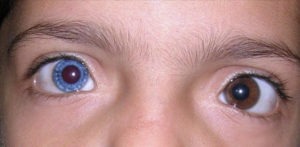
- Heterochromia iridis — complete heterochromia (one iris a different color from the other) or, more commonly, sectoral or partial heterochromia within a single iris, discussed further in this site’s dedicated coverage of iris heterochromia as a general finding, and one of the more visually striking and diagnostically useful clues to the syndrome
- Bright blue irides, in some patients, from hypopigmentation of both irises together rather than a color difference between them
- Dystopia canthorum — lateral displacement of the inner canthi with the puncta positioned more laterally than normal, giving a widened appearance to the space between the eyes even though the bony interorbital distance is actually normal (a distinction from true hypertelorism), present in type I but not type II
- Synophrys (medial fusion of the eyebrows) and a broad, high nasal root, contributing to the overall characteristic facial appearance
All-fit Slit-Lamp Adapter
Record and share exactly what you see at the slit lamp. One adapter fits any slit lamp or surgical microscope — and any smartphone.
From Choroida — the team behind this siteSystemic Findings
- Sensorineural hearing loss — the most functionally significant feature of the syndrome, present in a substantial proportion of affected individuals, ranging from mild to profound and from unilateral to bilateral
- A white forelock (a patch of prematurely white or gray hair at the front hairline) — a classically described and visually distinctive finding, though not present in every affected individual
- Patchy skin hypopigmentation (leukoderma)
- Premature graying of the hair more broadly, beyond just the classic forelock
- In type III: upper limb musculoskeletal abnormalities
- In type IV: Hirschsprung disease (congenital absence of ganglion cells in the distal colon, causing severe constipation and bowel obstruction), a serious associated finding that changes the urgency and nature of the required workup
Differential Diagnosis
- Piebaldism — an isolated pigmentary condition (white forelock, patchy skin depigmentation) without the hearing loss or characteristic dystopia canthorum of Waardenburg syndrome, caused by mutations in a different gene (KIT)
- Isolated congenital sensorineural hearing loss without associated pigmentary or craniofacial findings — the ocular and pigmentary findings of Waardenburg syndrome are precisely what distinguishes it from non-syndromic hearing loss, and their presence should prompt genetic evaluation specifically for this syndrome
- Tietz syndrome — a related but distinct condition combining deafness with more generalized albinism-like hypopigmentation, from a different MITF mutation than typical Waardenburg type II
Diagnostic Evaluation
Audiologic testing is essential in any child with suspected or confirmed Waardenburg syndrome, given how significantly hearing loss affects development if not identified and addressed early.
This is the single most clinically important step once the syndrome is suspected on the basis of its more visible pigmentary and craniofacial features, and should not be delayed while awaiting genetic confirmation.
Genetic testing confirms the specific subtype and causative mutation, useful both for prognostic counseling (regarding the risk of associated Hirschsprung disease in suspected type IV) and for family counseling given the syndrome’s autosomal dominant inheritance in most forms.
Management
There is no treatment for the underlying genetic condition.
Management is centered on early identification and treatment of hearing loss (hearing aids or cochlear implantation, depending on severity), which has a major impact on speech and language development if addressed promptly.
The ocular pigmentary findings themselves generally require no specific treatment, because they are cosmetic rather than functionally visually significant in the great majority of cases.
However, regular ophthalmic follow-up as part of routine pediatric eye care remains appropriate, including standard refraction and screening for any coexisting, unrelated ocular conditions.
In suspected type IV disease, evaluation for Hirschsprung disease is essential given its potential severity.
Genetic counseling for the family addresses recurrence risk for future children, given the syndrome’s predominantly autosomal dominant inheritance pattern.
Because expressivity varies considerably even within the same family, one affected parent may have only subtle pigmentary findings while their child has more pronounced hearing loss or other features, which is worth explaining clearly so families understand why severity can differ so much between relatives who carry the same underlying mutation.


Document what you see
Two smartphone imaging tools built for everyday clinic use — one for the slit lamp, one for the fundus.
From Choroida — the team behind this siteReferences
- Read AP, Newton VE. Waardenburg syndrome. Journal of Medical Genetics.
- Pingault V, Ente D, Dastot-Le Moal F, et al. Review and update of mutations causing Waardenburg syndrome. Human Mutation.
- American Academy of Ophthalmology. Basic and Clinical Science Course, Section 6: Pediatric Ophthalmology and Strabismus.
