
Chagas disease is caused by the protozoan parasite Trypanosoma cruzi and transmitted by triatomine (“kissing”) bugs.
When the parasite enters through the conjunctiva or periocular skin, it produces a recognizable eponymous sign in tropical medicine.
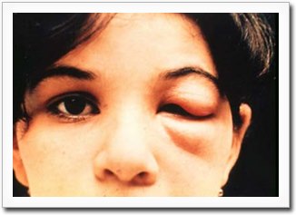
This is Romana’s sign: a unilateral, usually painless swelling of the eyelids and periorbital tissue.
Named after Cecilio Romaña, the Argentine physician who described it in 1935, the sign remains clinically relevant today.
This applies wherever Chagas disease is endemic, largely across Latin America.
However, rising population movement has made it a consideration in non-endemic countries as well.
The finding matters because it identifies the site of parasite entry during the acute phase.
At this point, the disease is far more treatable than once it progresses.
Later, it can reach the chronic cardiac or gastrointestinal complications that make Chagas disease so dangerous.
Mechanism
Infection typically occurs when triatomine bug feces are deposited near a bite wound on the skin.
These are then inadvertently rubbed into the eye or an area of broken skin.
The insect itself does not inject the parasite through its bite.
This is why the periocular route of entry is so mechanistically direct and so classically described.
The resulting local inflammatory reaction at the conjunctiva and periorbital tissue produces the characteristic unilateral swelling.
This occurs days to a few weeks after exposure.
The sign is a local reaction to parasite entry, rather than a systemic manifestation.
It appears only in patients infected through this specific route.
It is absent in most Chagas disease cases acquired through blood transfusion, congenital transmission, or contaminated food.
In these routes, the acute phase is often asymptomatic or produces only nonspecific fever.
Clinical Findings
- Unilateral, painless edema of the upper and lower eyelids on the side of inoculation
- Conjunctival injection and chemosis on the affected side
- Preauricular and submandibular lymphadenopathy, often palpable on the same side
- Associated acute-phase systemic findings — fever, malaise, and occasionally hepatosplenomegaly — in a minority of patients
The absence of pain and itching, together with the striking unilaterality, distinguishes Romana’s sign from more common causes of swelling.
These include allergic reaction, insect bite hypersensitivity, or preseptal cellulitis.
However, in practice these alternatives are still the more statistically likely explanation outside endemic areas.
They need to be actively excluded.
All-fit Slit-Lamp Adapter
Record and share exactly what you see at the slit lamp. One adapter fits any slit lamp or surgical microscope — and any smartphone.
From Choroida — the team behind this siteDifferential Diagnosis
- Preseptal or orbital cellulitis — usually painful, with erythema and warmth, and a source of local infection
- Allergic contact dermatitis or insect bite reaction — typically itchy, sometimes bilateral, with a clear exposure history
- Angioedema — can be unilateral but usually evolves faster (hours) and may involve other facial or mucosal sites
- Dacryoadenitis or dacryocystitis — localized to the lacrimal gland or sac region rather than diffuse periorbital swelling
A relevant history includes residence in or travel to an endemic region.
It also includes exposure to rural or peri-domestic housing where triatomine bugs live.
This history, together with the painless, unilateral quality of the swelling, should prompt specific consideration of Chagas disease.
It should not just be treated as a routine periorbital infection.
Diagnosis
During the acute phase, direct visualization of trypomastigotes on peripheral blood smear confirms the diagnosis.
PCR-based detection of parasite DNA also works, while parasitemia is still high enough to detect.
Serologic testing is used for chronic-phase diagnosis, once parasitemia has dropped below the detection threshold of direct methods.
Most national guidelines require at least two different assays for confirmation.
Management
Antitrypanosomal treatment — benznidazole or nifurtimox — is most effective when started during the acute phase.
This is precisely why recognizing Romana’s sign and other acute presentations promptly matters clinically.
Efficacy declines, though treatment is still often recommended, as the disease moves into the chronic phase.
The periorbital swelling itself resolves as the acute infection is treated and typically leaves no lasting ocular sequelae.
Beyond the acute presentation, the real long-term stakes of Chagas disease are cardiac (chronic Chagas cardiomyopathy) and gastrointestinal (megaesophagus, megacolon).
This is why any patient with a confirmed diagnosis needs baseline and ongoing screening for these complications.
This holds regardless of how mild the initial ocular presentation was.


Document what you see
Two smartphone imaging tools built for everyday clinic use — one for the slit lamp, one for the fundus.
From Choroida — the team behind this siteReferences
- Rassi A Jr, Rassi A, Marin-Neto JA. Chagas disease. Lancet.
- Pérez-Molina JA, Molina I. Chagas disease. Lancet.
- World Health Organization. Chagas disease (American trypanosomiasis) fact sheet.
- Bern C. Chagas’ disease. New England Journal of Medicine.
