
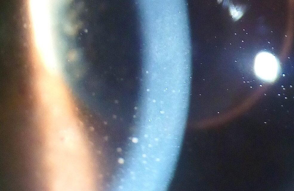
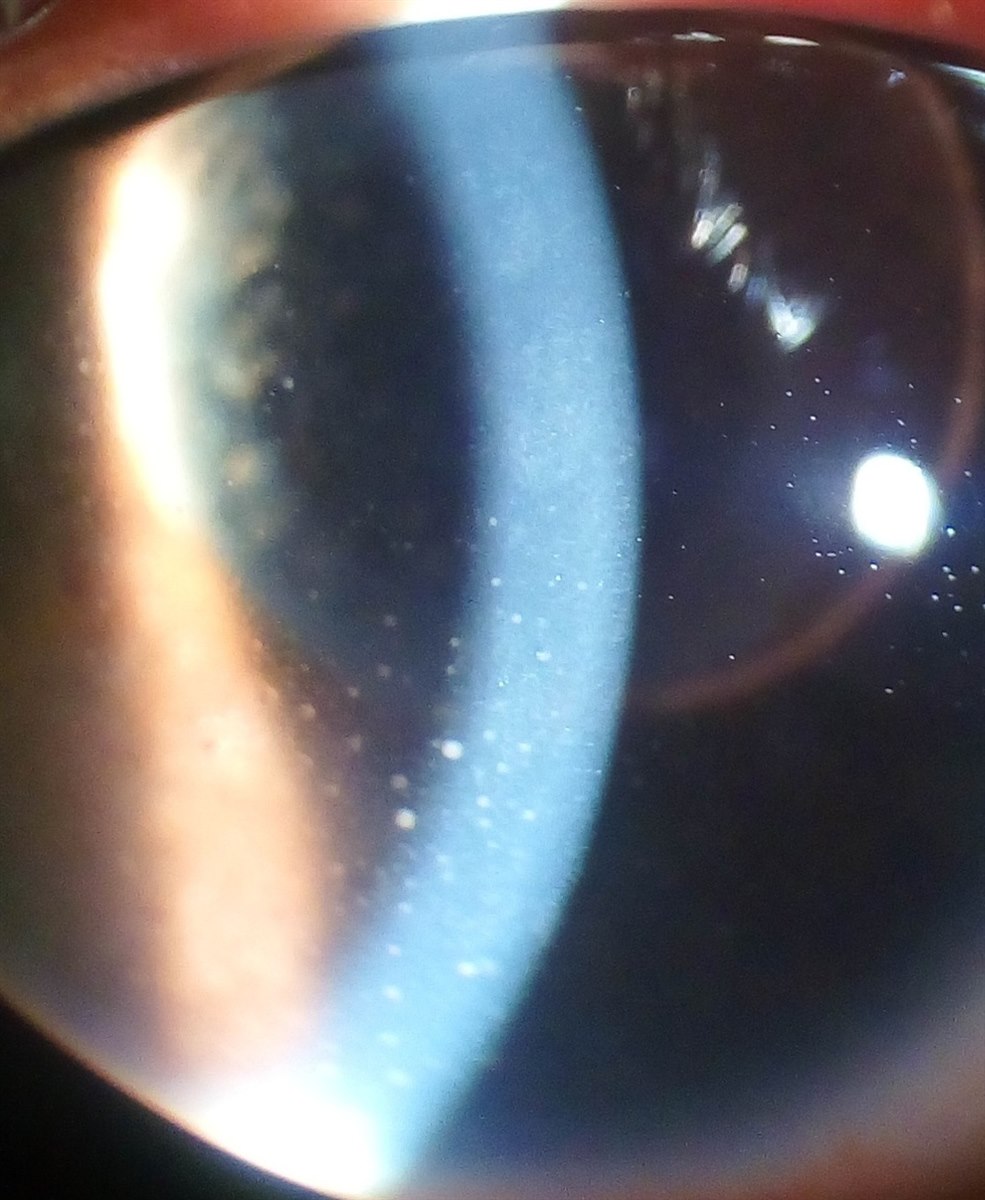
Keratic precipitates (KPs) are clumps of inflammatory cells deposited on the corneal endothelium, and they are one of the most useful signs in the entire uveitis exam, because their morphology alone can meaningfully narrow the differential before a single lab test comes back.
Aqueous convection currents carry cells shed into the anterior chamber during inflammation toward the corneal endothelium, where they settle, classically in a triangular distribution low and central, the so-called Arlt’s triangle, though the pattern varies with the underlying disease.
Learning to read KP morphology at the slit lamp is genuinely worth the effort, because granulomatous versus non-granulomatous appearance is one of the first branch points in working up any new anterior uveitis.
Morphologic Types
- Fine (non-granulomatous) KPs — small, white, dust-like deposits, typically composed of lymphocytes; associated with non-granulomatous uveitis such as HLA-B27-associated acute anterior uveitis, idiopathic uveitis, and many viral causes
- Mutton-fat (granulomatous) KPs — large, greasy-appearing, yellow-white deposits composed mainly of epithelioid cells and macrophages; classically associated with sarcoidosis, tuberculosis, sympathetic ophthalmia, and Vogt-Koyanagi-Harada disease
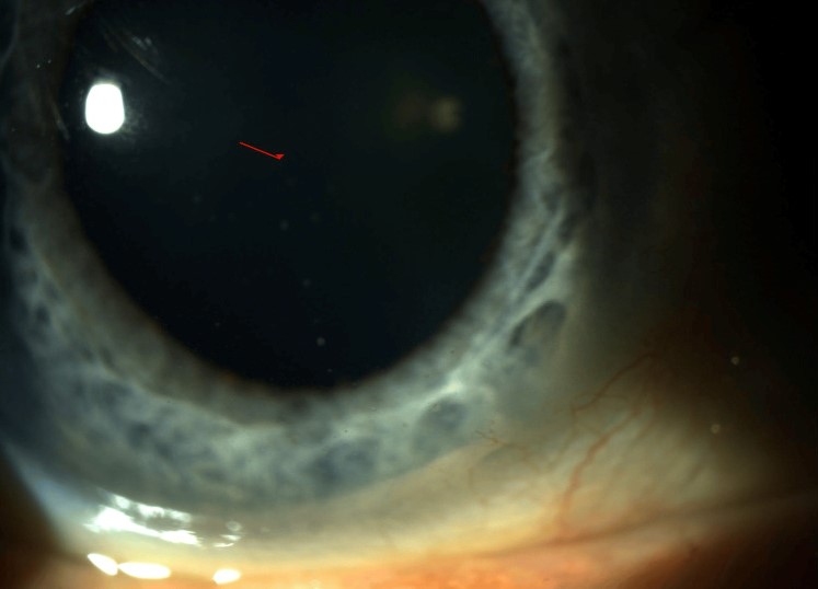
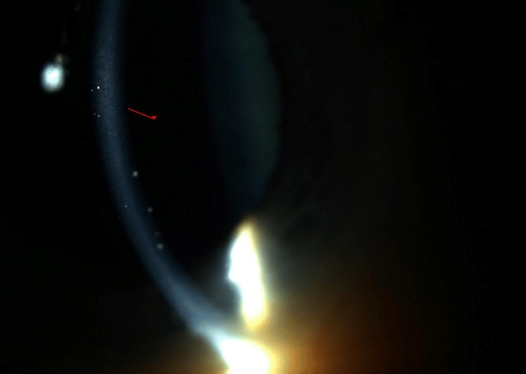
- Stellate KPs — small, star-shaped deposits with fine radiating processes, diffusely distributed across the endothelium rather than confined to Arlt’s triangle; the classic pattern in Fuchs heterochromic iridocyclitis
- Old (pigmented) KPs — inactive deposits that have become pigmented and fixed to the endothelium over time, indicating prior rather than necessarily active inflammation
What the Pattern Suggests
Granulomatous KPs shift the differential toward a specific, shorter list of conditions and typically prompt a targeted workup: chest imaging and ACE or lysozyme testing for sarcoidosis, tuberculin skin testing or interferon-gamma release assay for tuberculosis, and a careful history for prior ocular trauma or surgery if sympathetic ophthalmia is a concern.
Non-granulomatous KPs, by contrast, are compatible with a much broader range of causes and often do not warrant the same extensive systemic workup, especially in a first, unilateral, HLA-B27-compatible presentation.
The distribution matters alongside the morphology: diffuse, fine, stellate KPs without significant flare or synechiae — even in a quiet-looking eye — are a strong pointer toward Fuchs heterochromic iridocyclitis, a condition that is often misdiagnosed as “not uveitis” precisely because it lacks the dramatic redness and pain of other anterior uveitides.
Because morphology can shift somewhat as a single episode of uveitis evolves — older KPs within the same episode gradually taking on a more pigmented, less acute appearance — a single snapshot exam is sometimes less informative than tracking how the KP pattern changes, or fails to change, over serial visits.
All-fit Slit-Lamp Adapter
Record and share exactly what you see at the slit lamp. One adapter fits any slit lamp or surgical microscope — and any smartphone.
From Choroida — the team behind this siteAssociated Findings to Assess Alongside KPs
- Anterior chamber cell and flare grading, to establish overall inflammatory activity
- Posterior synechiae, indicating a more organized or longstanding inflammatory process
- Iris nodules — Koeppe (pupillary margin) or Busacca (iris stroma) nodules, more common with granulomatous disease
- Elevated intraocular pressure, common in both granulomatous uveitis and Fuchs heterochromic iridocyclitis, through different mechanisms
- Heterochromia, specifically in the context of Fuchs, where the affected iris is often (but not always) lighter rather than the fellow eye
Mimics of a True Stellate Pattern
- Herpetic anterior uveitis — can also produce diffuse, endothelium-wide KPs and elevated intraocular pressure, but typically with a more acute, painful presentation and a history of prior herpetic keratitis or characteristic corneal findings, distinguishing it from the quiet, chronic course of Fuchs heterochromic iridocyclitis
- Chronic, low-grade granulomatous uveitis — a diffuse rather than triangular distribution can superficially resemble a stellate pattern, but the individual deposits are mutton-fat rather than fine and star-shaped, and other supporting granulomatous features (Koeppe or Busacca nodules) are more often present
Correctly distinguishing a true stellate, Fuchs-type pattern from these mimics prevents both unnecessary aggressive anti-inflammatory treatment in Fuchs and, conversely, under-treatment of a genuinely more active granulomatous or herpetic process misread as the benign Fuchs pattern.
Evaluation
The workup for a new case of anterior uveitis is guided substantially by the KP morphology and the overall clinical picture — laterality, chronicity, associated systemic symptoms — rather than a reflexive, identical panel of tests for every patient.
A first episode of unilateral, non-granulomatous, acute anterior uveitis in an otherwise healthy adult, for example, is frequently managed with treatment and observation before extensive lab testing, if it resolves promptly, while granulomatous, bilateral, recurrent, or atypical presentations warrant a more thorough systemic evaluation from the outset.
This tiered approach — reserving the most extensive workup for the presentations most likely to actually have an identifiable, treatable systemic cause — avoids subjecting every patient with a first, straightforward uveitis episode to an expensive and often unrevealing battery of tests.
Management
Treatment targets the underlying uveitis rather than the KPs themselves: topical corticosteroids and cycloplegia for most anterior uveitis, with systemic treatment added when a specific systemic cause is identified or when the disease is severe, recurrent, or steroid-dependent.
Old, pigmented, inactive KPs from resolved past episodes generally do not require treatment on their own and can persist indefinitely as a marker of prior disease.
Fuchs heterochromic iridocyclitis in particular tends to respond poorly to corticosteroids and does not require the aggressive anti-inflammatory treatment used for other granulomatous or non-granulomatous uveitis, so correctly recognizing its distinctive stellate KP pattern actually changes management by avoiding unnecessary steroid exposure and its associated risks (cataract, glaucoma) in a condition that will not respond to it anyway.


Document what you see
Two smartphone imaging tools built for everyday clinic use — one for the slit lamp, one for the fundus.
From Choroida — the team behind this siteReferences
- Jabs DA, Nussenblatt RB, Rosenbaum JT. Standardization of uveitis nomenclature for reporting clinical data. American Journal of Ophthalmology.
- American Academy of Ophthalmology. Basic and Clinical Science Course, Section 9: Intraocular Inflammation and Uveitis.
- La Hey E, de Vries J, Langerhorst CT, et al. Treatment and prognosis of Fuchs’ heterochromic cyclitis. American Journal of Ophthalmology.
- Herbort CP, Rao NA, Mochizuki M. International criteria for the diagnosis of ocular sarcoidosis. Ocular Immunology and Inflammation.
