
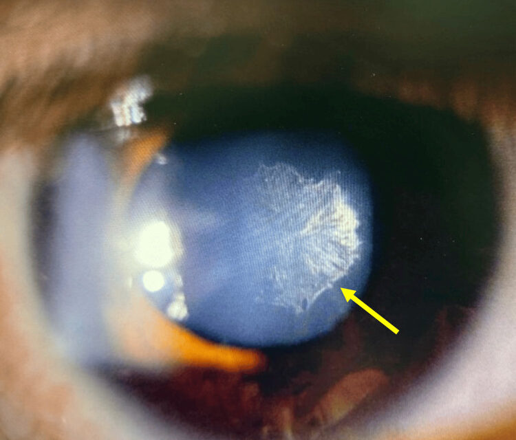
Posterior subcapsular cataract (PSC) is a distinct pattern of lens opacification that forms just in front of the posterior lens capsule, made up of granular, plaque-like deposits of aberrant lens epithelial cells that have migrated posteriorly and swollen (Wedl cells) rather than degenerated in the usual anterior-to-posterior direction of normal epithelial turnover.
It behaves differently from nuclear or cortical cataract in almost every practical respect — who gets it, how fast it progresses, and what symptoms it causes first — and recognizing this distinct pattern early can change both the diagnostic workup and the timing of any recommended treatment.
Because the opacity sits directly in the path of light converging toward the fovea, even a small PSC can cause visual symptoms disproportionate to its size, which is the single most useful clinical fact to remember about this cataract type.
Causes and Risk Factors
- Corticosteroid use — topical, systemic, or inhaled, and one of the most common identifiable causes, with risk roughly proportional to dose and duration
- Ocular inflammation — chronic uveitis of any cause is a major risk factor, independent of any steroid used to treat it
- Ionizing radiation exposure
- Diabetes mellitus
- High myopia
- Retinitis pigmentosa and other inherited retinal dystrophies
- Prior vitrectomy, which accelerates PSC formation through mechanisms that are not completely understood but are clinically well documented
Younger patients presenting with PSC should prompt specific questioning about steroid exposure — including inhaled steroids for asthma, which patients often do not think to mention — and screening for chronic intraocular inflammation, because PSC in a young adult is far less likely to be simple age-related change than nuclear or cortical cataract would be at the same age.
Clinical Presentation
Glare and difficulty in bright light are hallmark symptoms, because the opacity is most disruptive when the pupil is constricted (as it is in bright conditions), forcing light through the affected central zone rather than around it via the peripheral, clearer lens.
Patients often describe specific difficulty with oncoming headlights while driving at night and with reading in bright sunlight, a pattern distinct from the gradual, diffuse blur more typical of nuclear sclerosis.
Near vision is frequently affected earlier and more severely than distance vision, again related to pupillary constriction during near tasks, and visual acuity testing under standard exam-room lighting can actually underestimate how functionally impaired a patient is in bright, real-world conditions.
Because standard Snellen testing is performed under relatively dim, controlled lighting, specifically asking about glare and bright-light difficulty — rather than relying on the acuity number alone — is often what reveals the true functional impact of a PSC that otherwise looks modest on the chart, and a brief glare or contrast sensitivity test in the office can help document this discrepancy objectively.
All-fit Slit-Lamp Adapter
Record and share exactly what you see at the slit lamp. One adapter fits any slit lamp or surgical microscope — and any smartphone.
From Choroida — the team behind this siteExam Findings
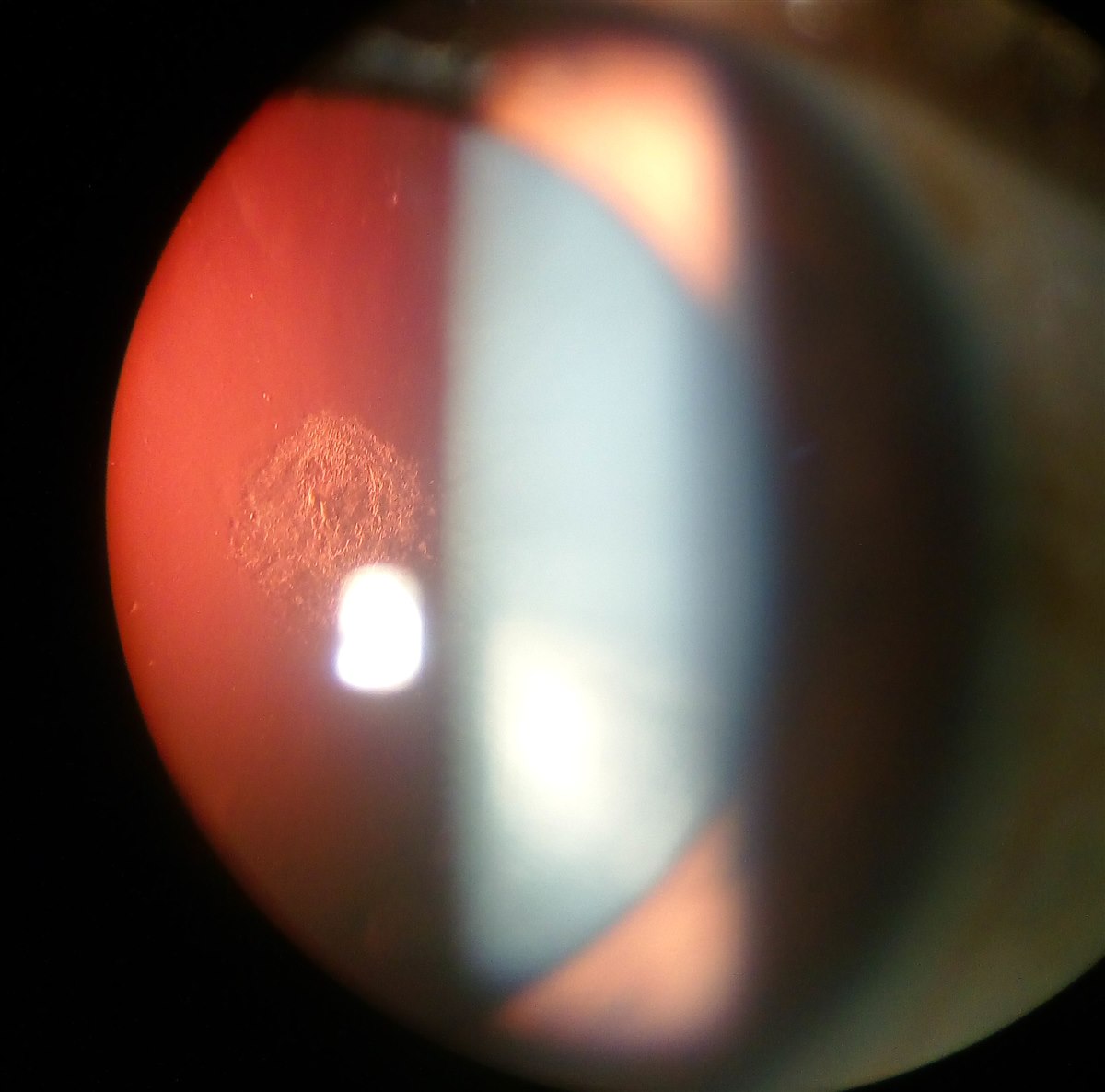
On direct or slit-lamp retroillumination against the red reflex, PSC appears as granular, sometimes iridescent plaque-like opacities concentrated at the posterior central lens capsule.
Progression tends to be faster than nuclear or cortical cataract: PSC can advance to visually significant levels over months in high-risk settings such as ongoing high-dose steroid use or active uveitis, rather than the years typically associated with age-related nuclear change.
A dilated exam is important not only to assess the cataract itself but to rule out the posterior segment disease (uveitis, retinitis pigmentosa) that may be driving it, because treating the cataract without addressing an active underlying cause invites recurrence in a fellow eye or after surgery in the pseudophakic capsule.
When active uveitis is present, quiet, well-controlled inflammation at the time of any planned cataract surgery is generally the goal, since operating on an actively inflamed eye substantially raises the risk of postoperative complications.
Differential Diagnosis
- Nuclear sclerotic cataract — central, yellow-brown, gradual myopic shift, distinct symptom pattern (worse for distance, better for near initially)
- Cortical cataract — peripheral spoke-like opacities, different glare pattern, slower functional impact until late
- Posterior capsule opacification (Elschnig pearls) — a similar-appearing finding but occurring after cataract surgery on a pseudophakic capsule, not a native lens finding
- Retained cortical material after prior surgery — relevant only in a pseudophakic or recently operated eye
Management
Addressing a reversible driver — tapering or replacing a chronic topical steroid where clinically feasible, or achieving better control of underlying uveitis — can slow progression, though established PSC opacity generally does not resolve once formed.
Because glare symptoms can be disabling even with preserved Snellen acuity in standard lighting, the threshold for surgical referral is often lower for PSC than for other cataract types; visual acuity alone can understate how much the cataract is limiting the patient.
Cataract surgery is effective for PSC, as it is for other cataract types, though patients with an underlying inflammatory or steroid-dependent cause need careful perioperative control of inflammation to reduce the risk of postoperative complications and posterior capsule opacification recurring on the new intraocular lens.
Patients with a steroid-related PSC also deserve counseling that the underlying condition requiring steroid treatment often still needs ongoing management, and that cataract surgery addresses the lens opacity itself without resolving whatever systemic or ocular disease made steroid treatment necessary in the first place, so continued coordination with whichever specialist prescribed the steroid remains important after eye surgery is complete.


Document what you see
Two smartphone imaging tools built for everyday clinic use — one for the slit lamp, one for the fundus.
From Choroida — the team behind this siteReferences
- Harding JJ, Egerton M, Harding RS. Risk factors for cataract in Oxfordshire: diabetes, peripheral neuropathy, myopia, glaucoma and diarrhoea. Acta Ophthalmologica.
- Chylack LT Jr. Mechanisms of senile cataract formation. Ophthalmology.
- Urban RC Jr, Cotlier E. Corticosteroid-induced cataracts. Survey of Ophthalmology.
- American Academy of Ophthalmology. Basic and Clinical Science Course, Section 11: Lens and Cataract.
