
CASE REPORT
A 42-year-old male presented to the ophthalmology clinic with chief complaints of blurred vision and floaters in both eyes.

His medical history revealed no significant systemic illnesses, but his occupational history included frequent exposure to dust and soil.
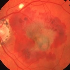
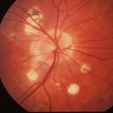
Examination findings indicated visual acuity of 20/40 in the right eye and 20/50 in the left eye, mild conjunctival injection in both eyes, clear corneas with no evidence of active inflammation, and characteristic fundus findings such as bilateral peripapillary atrophy and multiple chorioretinal scars scattered throughout the macular region.
Fluorescein angiography confirmed the absence of active leakage or neovascularization.
Diagnostic testing included documented chorioretinal scars through fundus photography, a negative chest X-ray for acute pulmonary histoplasmosis infection, and the absence of histopathological examination due to the patient’s asymptomatic systemic status.
Based on these findings, the patient was diagnosed with Presumed Ocular Histoplasmosis Syndrome (POHS).
DISEASE entity
Presumed ocular histoplasmosis syndrome (POHS) occurs secondary to infection with the yeast form of Histoplasma capsulatum.
The disease is characterized by atrophic chorioretinal scars, peripapillary atrophy (PPA), and the absence of vitritis. POHS is asymptomatic until choroidal neovascularization (CNV) or sequelae such as disciform scars develop.
There is considerable controversy over the cause of Presumed ocular histoplasmosis syndrome (POHS). Classically, POHS is caused by infection with the yeast form of H. capsulatum, which is a dimorphic fungus that lives in the soil.
It is endemic to states that contain the Ohio and Mississippi river valleys. Although distributed worldwide, POHS has only been reported in a few non-U.S. countries including Mexico, India, the United Kingdom, and the Netherlands.
It is carried on the feathers of chickens, pigeons, and blackbirds as well as in the droppings from infected bats.
Infection in humans occurs after inhalation of the spore or conidia form, classically associated with cave exploration and exposure to infected bats as well as birds, followed by hematogenous spread of the organism to the choroid.
The link between H. capsulatum and POHS comes from epidemiological studies where POHS was linked to a positive histoplasmin skin antigen test.
However, a study in the Netherlands found that all patients with clinical POHS were histoplasmin skin antigen test negative.
POHS is different from “Disseminated Histoplasmosis” which is the exudative/productive H. capsulatum infection of ocular tissues, seen in immunocompromised adults or infants.
Since Presumed ocular histoplasmosis syndrome (POHS) is linked to HLA haplotypes DRw2 and B7, some hypothesize that it could also represent an autoimmune inflammatory reaction triggered by certain organisms, including H. capsulatum.
HLA DRw2 has been found in higher concentrations in patients with disciform and peripheral scars compared to the normal population.
Similarly, nearly 78% of patients with clinical POHS and macular disciform scars in at least one eye are positive for HLA-B7. The absence of vitritis in the clinical criteria for POHS also supports an inflammatory rather than an active infectious etiology.

The diagnosis of Presumed ocular histoplasmosis syndrome (POHS) is based upon a fundoscopic exam for the four cardinal features and fluorescein angiography for the characterization of CNV.
Fundus Explorer Pro
Photograph the retinal findings described here with the phone already in your pocket — 22 D optics and built-in illumination in one handheld unit.
From Choroida — the team behind this siteSigns of Presumed ocular histoplasmosis syndrome (POHS)
Three characteristic findings of POHS are typically found on fundoscopic exams: multiple white atrophic chorioretinal scars or histo spots, PPA, and the absence of vitritis.
These findings may or may not be associated with CNV, which often appears as greenish-yellow subretinal discoloration with a surrounding pigmented ring at the macula.
Confluent atrophic chorioretinal scars in a linear or curvilinear pattern of variable length, width, and pigmentation can be found in the mid-periphery in approximately 5% of patients. In advanced cases, CNV can progress to a disciform scar with subretinal fibrovascular tissue. POHS changes can be found bilaterally in up to 60% of cases.
Presumed ocular histoplasmosis syndrome (POHS) Symptoms
Prior to CNV development, Presumed ocular histoplasmosis syndrome (POHS) was asymptomatic. POHS often presents with the classic symptoms of CNV, including painless vision loss, metamorphopsia, blurred central vision, and central or paracentral scotomas.
Presumed ocular histoplasmosis syndrome (POHS) MANAGEMENT
Presumed ocular histoplasmosis syndrome (POHS) without CNV is observed and counseling for monitoring of CNV. Because POHS is not truly an infectious process, early attempts at anti-fungal therapy yielded no benefit.
Treatment is generally guided toward early detection of CNV via Amsler grid screening and routine dilated fundoscopic exams. Once CNV develops, the approach is similar to the treatment of AMD.
Read more about management strategies


Document what you see
Two smartphone imaging tools built for everyday clinic use — one for the slit lamp, one for the fundus.
From Choroida — the team behind this siteREFERENCES
- Ophthalmology, A.A.o. in Basic and Clinical Sciences Course (Lifelong Education for the Ophthalmologist, San Fransisco, CA, 2006)
- Oliver, A., Ciulla, T.A. & Comer, G.M. 2005. New and classic insights into presumed ocular histoplasmosis syndrome and its treatment. Curr Opin Ophthalmol 16: 160-5.
- Prasad, A.G. & Van Gelder, R.N. 2005. Presumed ocular histoplasmosis syndrome. Curr Opin Ophthalmol 16: 364-8.
- Ongkosuwito, J.V., Kortbeek, L.M., Van der Lelij, A., Molicka, E., Kijlstra, A., de Smet, M.D. & Suttorp-Schulten, M.S. 1999. Aetiological study of the presumed ocular histoplasmosis syndrome in the Netherlands. Br J Ophthalmol 83: 535-9.
- Giles, C.L. & Falls, H.F. 1961. Further evaluation of amphotericin-B therapy in presumptive histoplasmosis chorioretinitis. Am J Ophthalmol 51: 588-98.
