Bleb grading is the structured scoring of a filtering bleb’s area, height, vascularity and leakage after glaucoma surgery.
The Moorfields Bleb Grading System and the Indiana Bleb Appearance Grading Scale are the two most widely used methods. Both turn a slit lamp impression into numbers, so clinicians can compare findings across visits and examiners.
Why Bleb Grading Matters
The bleb is the visible end of the aqueous pathway created at surgery (see trabeculectomy). Its appearance changes most in the first three months, while wound healing decides the outcome. However, a note such as “good bleb” tells the next examiner very little.
How to Examine the Bleb
Ask the patient to look down so the superior conjunctiva is fully exposed. Then use diffuse light for extent and vascularity, and a slit beam for height and wall thickness. Finally, apply fluorescein for a Seidel test and measure intraocular pressure, because appearance alone never confirms function.
All-fit Slit-Lamp Adapter
Record and share exactly what you see at the slit lamp. One adapter fits any slit lamp or surgical microscope — and any smartphone.
From Choroida — the team behind this siteThe Moorfields Bleb Grading System
Wells and colleagues described this system in 2004, using a set of reference photographs. It grades central area, maximal area, height and vascularity, plus subconjunctival blood. The system also scores vascularity separately for the central bleb, the peripheral bleb and nonbleb conjunctiva.
- Area (central and maximal): 1 to 5 against reference images
- Height: 1 (flat) to 4 (high)
- Vascularity: 1 avascular, 2 normal, 3 mild, 4 moderate, 5 severe
The Indiana Scale for Bleb Grading
Cantor and colleagues published the Indiana scale in 2003, also based on standard slit lamp photographs. Each parameter receives a letter and number, so a full description fits in one line, for example H2 E3 V1 S0.
- Height: H0 flat to H3 high
- Extent: E0 under one clock hour to E3 four clock hours or more
- Vascularity: V0 avascular white, V1 avascular cystic, V2 mild, V3 moderate, V4 extensive
- Seidel: S0 none, S1 pinpoint leaks, S2 streaming within five seconds
Moorfields Versus Indiana Bleb Grading
A 2006 comparison by Wells and coauthors reported acceptable reproducibility for both systems. Moorfields gives finer vascular detail and separates central from total area, while Indiana is faster and scores leakage directly. Therefore, the choice often depends on whether a clinic needs speed or research detail.
What Vascularity Predicts
Early hyperemia is the most studied warning sign on serial examination, and dense, tortuous corkscrew vessels over the bleb suggest an active healing response and impending scarring. As a result, a red bleb in the first weeks often prompts more frequent topical steroids or an antimetabolite injection.
What Height and Extent Predict
A diffuse, moderately elevated bleb over several clock hours usually reflects good filtration. However, a flat bleb with rising pressure suggests scarring at the scleral flap or under the conjunctiva. A flat bleb with low pressure and a shallow chamber instead raises concern for overfiltration or a leak.
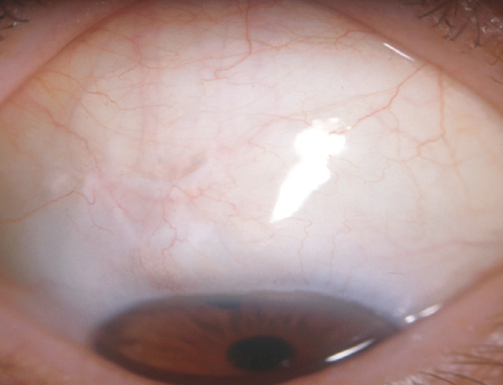
Encapsulated Blebs in Bleb Grading
An encapsulated bleb (Tenon cyst) is a tense, high, dome-shaped elevation with a thick wall and engorged vessels. It usually appears within the first few weeks to months, and pressure rises because aqueous cannot cross the capsule. Options include massage, aqueous suppressants and needling with an antimetabolite (see bleb needling).
Avascular Cystic Blebs
A thin, pale, cystic bleb often follows mitomycin C and may control pressure over the long term, but the thin wall predisposes to late leaks, hypotony and infection. Therefore, teach these patients the warning signs of blebitis and bleb-associated endophthalmitis.
Seidel Testing in Bleb Grading
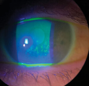
A positive test shows dark aqueous streaming through bright fluorescein, and pinpoint oozing may respond to conservative measures, while a brisk leak with hypotony usually needs surgical revision. In addition, always test a flat, low-pressure bleb, because slow leaks are easy to miss.
Imaging Beyond Clinical Bleb Grading
In addition, anterior segment OCT shows the bleb’s internal structure in cross section. Functioning blebs often show a hyporeflective interior, a visible fluid channel and epithelial microcysts. Failing blebs, by contrast, tend to look dense and highly reflective.
Using Bleb Grading at Follow-Up
Grade at each early postoperative visit with the same system, ideally with photographs. Then compare scores in sequence instead of judging one visit in isolation. A rising vascularity grade with falling height is therefore more concerning than either change alone.
Discussing the Bleb With Patients
Explain that the bleb is a small drainage reservoir hidden under the upper lid. Also advise patients to avoid rubbing and to report redness, discharge, pain or blurred vision at once. In addition, warn that the bleb may need needling or other procedures later.
Pitfalls to Avoid
- Do not equate a large bleb with a healthy bleb
- Never grade a bleb without measuring intraocular pressure
- Avoid mixing Moorfields and Indiana scores in one record
- Remember that steroids and antimetabolites can also lower vascularity grades


Document what you see
Two smartphone imaging tools built for everyday clinic use — one for the slit lamp, one for the fundus.
From Choroida — the team behind this siteReferences
- Cantor LB, Mantravadi A, WuDunn D, Swamynathan K, Cortes A. Morphologic classification of filtering blebs after glaucoma filtration surgery: the Indiana Bleb Appearance Grading Scale. J Glaucoma. 2003;12:266-271.
- Wells AP, Crowston JG, Marks J, et al. A pilot study of a system for grading of drainage blebs after glaucoma surgery. J Glaucoma. 2004;13:454-460.
- Wells AP, Ashraff NN, Hall RC, Purdie G. Comparison of two clinical bleb grading systems. Ophthalmology. 2006;113:77-83.
- American Academy of Ophthalmology. Basic and Clinical Science Course, Section 10: Glaucoma. San Francisco: AAO; 2023.
- Salmon JF. Kanski’s Clinical Ophthalmology: A Systematic Approach. 9th ed. Elsevier; 2020.

Writes and reviews the clinical articles on Educate. Every post is checked against peer-reviewed sources before it goes live.