Corneal ulcer grading sorts infectious keratitis by size, depth, location and intraocular reaction.
No single scale is universally validated, and cut-offs differ between centers. However, most schemes share the same features, and those features guide referral, culture and drug choice.
What Corneal Ulcer Grading Measures
A grade estimates two risks: losing vision and losing the globe. Size and depth reflect stromal burden, while location predicts the visual cost of a scar, and the anterior chamber reaction shows how intense the inflammation has become.
Size and Depth in Corneal Ulcer Grading
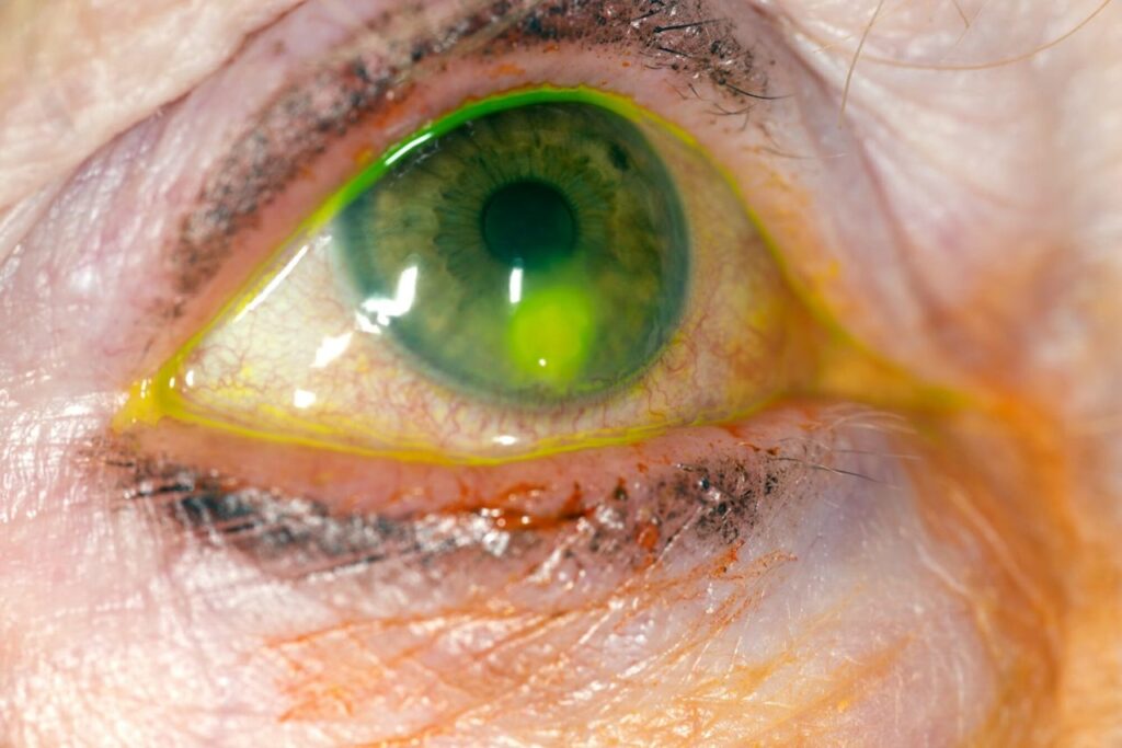
First, measure the epithelial defect and the stromal infiltrate separately with the slit beam, and record the longest diameter and its perpendicular, because the two lesions often differ. Then use a narrow, oblique beam to place the infiltrate in the anterior, mid or posterior stroma, and anterior segment OCT can also document thinning (see corneal pachymetry).
All-fit Slit-Lamp Adapter
Record and share exactly what you see at the slit lamp. One adapter fits any slit lamp or surgical microscope — and any smartphone.
From Choroida — the team behind this siteLocation and Anterior Chamber Reaction
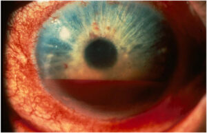
Central ulcers threaten acuity even when small, because any scar sits on the visual axis, while limbal lesions can spread to the sclera, where topical therapy works poorly. Hypopyon usually signals severe inflammation, although it is often sterile in bacterial keratitis, and fungal keratitis needs more caution, because organisms can penetrate Descemet membrane.
The 1-2-3 Rule in Corneal Ulcer Grading
Vital and colleagues proposed a simple severity rule for triage, which flags 1+ cells, an infiltrate of 2 mm or more, or a lesion within 3 mm of the center. A flagged ulcer should prompt culture and consideration of referral, but as a screening tool the rule favors caution, so some flagged ulcers will still do well.
Corneal Ulcer Grading: Mild, Moderate, Severe
The scale below is a practical summary, and local protocols may use different thresholds.
- Mild: infiltrate under 2 mm, superficial, away from the visual axis, with minimal reaction
- Moderate: infiltrate about 2 to 5 mm, up to about half depth, with moderate reaction
- Severe: infiltrate over 5 mm, deeper than half, marked thinning, hypopyon or scleral extension
In practice, let the worst single feature set the grade.
Features That Raise the Grade
Some findings raise concern even when the ulcer looks small.
- Satellite lesions or feathery margins suggesting fungal infection
- Ring infiltrate or severe pain suggesting Acanthamoeba
- A corneal graft, recent ocular surgery or an only eye
- No improvement after 48 hours of treatment
Who to Refer After Corneal Ulcer Grading
Refer moderate and severe ulcers urgently to a cornea service, and similarly refer any central ulcer, suspected atypical organism or failure of first-line therapy. A small peripheral ulcer in a reliable patient can often stay with the treating clinician, provided review is close.
Risk profile also lowers the threshold (see microbial keratitis risk factors). Perforation or impending perforation needs same-day specialist care, and the next step is to protect the eye with a rigid shield and avoid pressure on the globe.
Cultures and Severity
The AAO guideline supports culture for central, large or stromal ulcers, so take scrapings before starting antimicrobials when this causes no meaningful delay. A negative culture does not exclude infection, because earlier antibiotics reduce yield.
Treatment by Severity
Topical fluoroquinolone monotherapy is a common empirical choice for small peripheral bacterial ulcers, while the patient must stop contact lens wear and return within 24 to 48 hours. Severe ulcers often receive fortified antibiotics, with loading doses and then hourly drops.
For filamentous fungal ulcers, MUTT I favored natamycin over voriconazole. Finally, perforation may need glue or keratoplasty, because drops cannot restore lost tissue.
Steroids and Severity
SCUT found no overall benefit from adjunctive steroids in bacterial ulcers at three months, although subgroup data suggested possible benefit in eyes with very poor baseline vision (see the SCUT trial). Avoid steroids in suspected fungal disease.
Corneal Ulcer Grading at Follow-Up
A grade can change, so reassess it at every visit, and improvement includes less pain, blunted margins, a shrinking defect and a quieter chamber. By contrast, enlarging infiltrate, new thinning or rising hypopyon should trigger escalation and repeat culture, and consider drug toxicity when the infiltrate improves while the epithelium fails to heal.
Explaining the Grade to Patients
Explain that the grade reflects current risk and may change within days, then describe the drop schedule clearly, including night doses when required. Warn that healing can still leave a scar and reduced vision.
Pitfalls to Avoid
- Do not grade by the epithelial defect alone
- Never dismiss a small central ulcer as mild
- Avoid steroids before excluding fungal and herpetic disease


Document what you see
Two smartphone imaging tools built for everyday clinic use — one for the slit lamp, one for the fundus.
From Choroida — the team behind this siteReferences
- Vital MC, Belloso M, Prager TC, Lanier JD. Classifying the severity of corneal ulcers by using the “1, 2, 3” rule. Cornea. 2007;26:16-20.
- Lin A, Rhee MK, Akpek EK, et al. Bacterial Keratitis Preferred Practice Pattern. Ophthalmology. 2019;126:P1-P55.
- Srinivasan M, Mascarenhas J, Rajaraman R, et al. Corticosteroids for bacterial keratitis: the Steroids for Corneal Ulcers Trial (SCUT). Arch Ophthalmol. 2012;130:143-150.
- Prajna NV, Krishnan T, Mascarenhas J, et al. The Mycotic Ulcer Treatment Trial: a randomized trial comparing natamycin vs voriconazole. JAMA Ophthalmol. 2013;131:422-429.
- Austin A, Lietman T, Rose-Nussbaumer J. Update on the management of infectious keratitis. Ophthalmology. 2017;124:1678-1689.
Test yourself
A few questions straight from this article.
-
Which two risks does a corneal ulcer severity grade estimate?
A grade estimates the risk of losing vision and of losing the globe; size and depth reflect stromal burden, location the visual cost and the anterior chamber the inflammation. -
Why does a limbal location raise concern when grading a corneal ulcer?
Central ulcers threaten acuity even when small, while limbal lesions can spread to the sclera, where topical therapy works poorly. -
In the 1-2-3 rule for corneal ulcer triage, what does the '2' flag?
The 1-2-3 rule of Vital and colleagues flags 1+ anterior chamber cells, an infiltrate of 2 mm or more, or a lesion within 3 mm of the center, prompting culture and consideration of referral. -
On a practical mild-to-severe corneal ulcer scale, which infiltrate size marks a severe ulcer?
A severe ulcer has an infiltrate over 5 mm, deeper than half thickness, marked thinning, hypopyon or scleral extension; moderate is about 2 to 5 mm. -
When corneal ulcer features fall into different severity grades, which rule sets the overall grade?
In practice the worst single feature sets the grade, and the epithelial defect alone should never be used to grade an ulcer. -
Which finding raises a corneal ulcer's grade even when the ulcer looks small?
Features that raise the grade include no improvement after 48 hours, a corneal graft, recent surgery or an only eye, and signs suggesting fungal or Acanthamoeba infection. -
Which corneal ulcer can often stay with the treating clinician rather than be referred to a cornea service?
Moderate and severe, central, atypical or non-responding ulcers should be referred urgently; a small peripheral ulcer in a reliable patient can often stay local with close review. -
A corneal ulcer shows impending perforation. What should be done while arranging same-day specialist care?
Perforation or impending perforation needs same-day specialist care, and the eye should be protected with a rigid shield while avoiding pressure on the globe. -
What is a common empirical regimen for a small peripheral bacterial corneal ulcer?
Fluoroquinolone monotherapy is a common empirical choice for small peripheral bacterial ulcers, with contact lens wear stopped and review within 24 to 48 hours; severe ulcers often get fortified antibiotics. -
During corneal ulcer follow-up, the infiltrate improves but the epithelium fails to heal. What should be considered?
Drug toxicity should be considered when the infiltrate improves while the epithelium fails to heal; enlarging infiltrate, new thinning or rising hypopyon instead trigger escalation and repeat culture.

Writes and reviews the clinical articles on Educate. Every post is checked against peer-reviewed sources before it goes live.