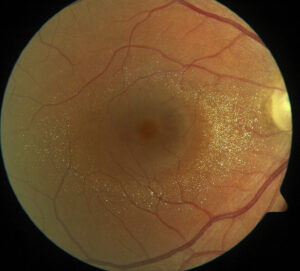
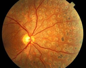
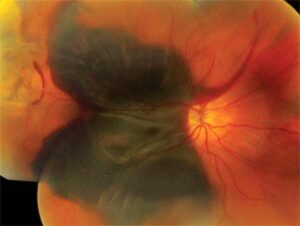
The Amsler grid is a simple chart of horizontal and vertical lines that tests the central 10 degrees of the visual field.
Marc Amsler introduced it in the 1940s, and clinicians still use it to detect metamorphopsia and central scotomas.
It is cheap and quick, but it has limits that every user should understand.
How to Perform the Amsler Grid Test
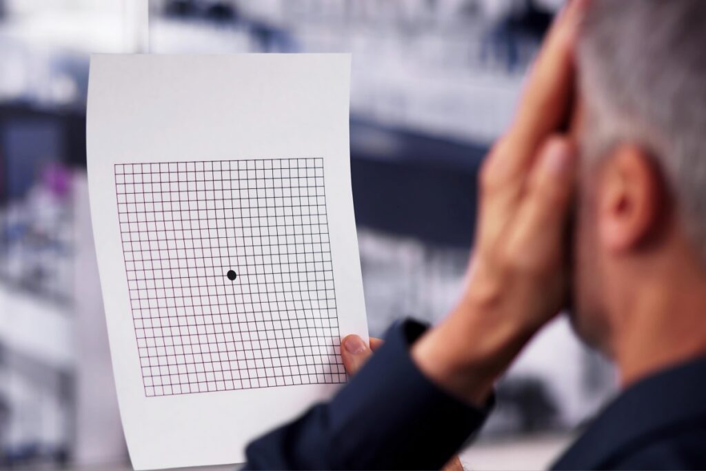
The standard grid is a 10 cm square with 5 mm boxes, viewed from about 30 cm.
The patient wears reading glasses, covers one eye, and fixates on the central dot.
Ask the patient whether any lines look wavy, missing, or blurred, and whether the corners are visible.
Then ask the patient to draw any abnormal area on the chart, and test the other eye (see retinal imaging).
Reading the Amsler Grid Results
Wavy or bent lines indicate metamorphopsia, which may arise from fluid, membranes, or traction.
A blank or gray area suggests a scotoma, such as in macular scars or geographic atrophy.
Missing corners may signal a field defect, and a central distortion can point to macular disease instead of optic nerve disease.
Record the findings on the grid and in the chart, so that follow-up changes are clear.
Fundus Explorer Pro
Photograph the retinal findings described here with the phone already in your pocket — 22 D optics and built-in illumination in one handheld unit.
From Choroida — the team behind this siteUses of the Amsler Grid
- Detecting new neovascular membranes in patients with AMD
- Monitoring macular holes, epiretinal membranes, and central serous disease
- Screening for toxic maculopathy in selected cases
- Checking symptoms after retinal surgery
Fine and colleagues reported that distortion was one of the earliest symptoms of a new neovascular membrane.
Evidence and Limits of the Amsler Grid
Schuchard found that patients often fail to report scotomas on the grid, because the brain fills in missing areas.
Therefore, the grid may miss real defects, especially small ones.
The HOME study compared a preferential hyperacuity perimetry device with standard care and found earlier detection with the device.
These results show that the grid alone gives limited sensitivity for early disease (see age-related macular degeneration management).
Common Errors With the Amsler Grid
- Using the grid without the correct reading glasses
- Testing both eyes at once, so the better eye hides the problem
- Holding the grid at the wrong distance
- Failing to fixate on the central dot
Use the grid under good lighting and give clear instructions.
An Example Case
A 74-year-old man with intermediate AMD in his right eye checks an Amsler grid at home every morning.
One day he notices that the lines in the center of the grid look wavy, and the change is new.
He calls the clinic, and OCT the same week shows subretinal fluid from a new neovascular membrane.
He starts anti-VEGF treatment within days, and his acuity stays at 20/40.
Questions Patients Ask
Patients ask how often to check the grid, and daily checks give the best chance of catching change.
They also ask what to do if the lines change, and the answer is to call the clinic at once, not to wait for the next visit.
Because patients may forget, link the check to a daily habit such as reading the morning paper.
Pitfalls to Avoid
- Relying on the grid as the only monitoring tool in high-risk eyes
- Giving the chart without teaching how to use it
- Ignoring a patient’s report of new distortion because the grid looks normal
- Forgetting that the brain fills in small scotomas
Looking Ahead
Digital grids and smartphone applications may improve compliance.
Home OCT and preferential hyperacuity devices offer higher sensitivity for neovascular conversion.
However, the paper grid remains a free and accessible starting point.
Practical Points
Give each at-risk patient a printed grid and a short instruction sheet.
Then ask the patient to demonstrate the test at the next visit.
Also record any new distortion in the chart, since it may predict later changes.
Teaching Patients to Use the Grid
Demonstrate the test in the clinic and let the patient try it.
Then give clear rules for what to do when the lines change.
Because many patients fear the answer, reassure them that early reporting protects vision.
Also ask them to bring the grid to each visit.
Alternatives to the Amsler Grid
Digital tests, such as preferential hyperacuity perimetry and tablet applications, add sensitivity.
However, access and cost vary, so many clinics still rely on the paper grid.
Choose the tool that the patient will actually use.


Document what you see
Two smartphone imaging tools built for everyday clinic use — one for the slit lamp, one for the fundus.
From Choroida — the team behind this siteReferences
- Fine AM, Elman MJ, Ebert JE, et al. Earliest symptoms caused by neovascular membranes in the macula. Arch Ophthalmol. 1986;104:513-514.
- Schuchard RA. Validity and interpretation of Amsler grid reports. Arch Ophthalmol. 1993;111:776-780.
- Chew EY, Clemons TE, Bressler SB, et al. Randomized trial of a home monitoring system for early detection of choroidal neovascularization: Home Monitoring of the Eye (HOME) study. Ophthalmology. 2014;121:535-544.
- American Academy of Ophthalmology. Basic and Clinical Science Course, Section 12: Retina and Vitreous. AAO; current edition.
Test yourself
A few questions straight from this article.
-
Which portion of the visual field does the Amsler grid test?
The Amsler grid is a chart of horizontal and vertical lines that tests the central 10 degrees of the visual field, which is why it suits macular disease. -
At what distance is the standard 10 cm Amsler grid viewed?
The standard grid is a 10 cm square of 5 mm boxes held at about 30 cm, and holding it at the wrong distance is a common error. -
On Amsler grid testing, what do wavy or bent lines indicate?
Wavy or bent lines indicate metamorphopsia, which may arise from macular fluid, membranes or traction. -
Why do patients often fail to report scotomas on the Amsler grid?
Schuchard found that the brain fills in missing areas, so the grid can miss real defects, especially small ones. -
In the HOME study, which approach detected neovascular AMD earlier than standard care?
The HOME study compared a preferential hyperacuity perimetry device with standard care and found earlier detection with the device, showing the grid alone has limited sensitivity. -
Why must each eye be tested separately on the Amsler grid?
Testing both eyes at once lets the better eye mask a defect, so the patient covers one eye and fixates on the central dot. -
A patient with AMD notices new wavy lines on a home Amsler grid. What is the correct advice?
New distortion should prompt an immediate call to the clinic rather than waiting, because it may signal a new neovascular membrane. -
How often should at-risk patients check the Amsler grid at home?
Daily checks give the best chance of catching change, and linking the check to a daily habit helps patients remember. -
On the Amsler grid, central distortion points toward disease at which site?
A central distortion on the grid points to macular disease rather than optic nerve disease, while missing corners may signal a field defect. -
What correction should a patient wear for Amsler grid testing?
The patient wears reading glasses for the test; using the grid without the correct reading glasses is a common error.

Writes and reviews the clinical articles on Educate. Every post is checked against peer-reviewed sources before it goes live.