Simple myopic hemorrhage is a subretinal bleed in a highly myopic eye that occurs without choroidal neovascularization.
It usually marks the formation of a new lacquer crack in Bruch membrane. Because it mimics myopic CNV, the main task is to exclude a neovascular membrane.
What Simple Myopic Hemorrhage Is
As the globe elongates, Bruch membrane stretches and can split, and the break tears the adjacent choriocapillaris, so blood escapes into the subretinal space or outer retina. No new vessel forms, so the lesion has no exudative activity, and Ohno-Matsui and colleagues described this bleed as a sign of new lacquer crack formation.
Who Gets It
- High myopia with long axial length
- Younger adults, often younger than typical CNV patients
- Existing lacquer cracks or diffuse chorioretinal atrophy
- Posterior staphyloma
- Recent straining or minor trauma in some cases
Fundus Explorer Pro
Photograph the retinal findings described here with the phone already in your pocket — 22 D optics and built-in illumination in one handheld unit.
From Choroida — the team behind this siteSymptoms of Simple Myopic Hemorrhage
Patients report sudden central blur, a dark spot or distortion, and these symptoms match myopic CNV, so history alone cannot separate them. However, some patients recall heavy lifting or straining before onset, while pain and redness are absent.
Fundus Signs of Simple Myopic Hemorrhage
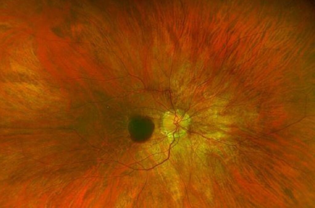
The bleed is usually small, round and deep red, at or near the fovea, and it lacks the gray membrane or pigment ring of active CNV. Hard exudate and retinal thickening are absent, and in addition, look for yellow linear lacquer cracks nearby, although the crack may stay hidden until the blood clears.
OCT in Simple Myopic Hemorrhage
OCT is the first test to order, and blood appears as hyperreflective material in the outer retina or subretinal space, with shadowing below. However, there is no dome-shaped hyperreflective lesion above the RPE with fuzzy borders, and subretinal and intraretinal fluid are also absent, which argues against active CNV.
OCTA and Ruling Out CNV
OCTA searches for flow in the outer retina and choriocapillaris slabs (see OCT angiography), and in simple hemorrhage, these slabs show no abnormal vascular network. However, dense blood can mask flow signal and hide a small membrane, so repeat OCTA after the blood thins if doubt remains.
Fluorescein and ICG Angiography
Fluorescein angiography shows blocked fluorescence without late leakage, while leakage beyond the blood border suggests CNV instead. Meanwhile, indocyanine green angiography can show the lacquer crack as a late linear hypofluorescent band.
Simple Myopic Hemorrhage Versus Myopic CNV
The two conditions share symptoms, age range and location, but CNV shows a subretinal hyperreflective lesion, leakage on fluorescein and flow on OCTA (see myopic choroidal neovascularization). Also consider idiopathic CNV, punctate inner choroidopathy and traumatic choroidal rupture, and when findings are equivocal, review within weeks instead of assuming a benign bleed.
Why Most Cases Resolve
The bleed comes from a single break that seals quickly, and because no leaking vessel feeds it, blood stops accumulating. As a result, the hemorrhage usually absorbs over weeks to a few months, and observation alone is appropriate once CNV is excluded.
Visual Prognosis After Simple Myopic Hemorrhage
Most patients regain good vision once the fovea clears, although a crack through the fovea may leave a permanent scotoma or mild distortion. In addition, outer retinal damage on OCT after absorption predicts a weaker recovery, and older eyes with background atrophy tend to recover less.
Why Anti-VEGF Is Not Indicated
Anti-VEGF drugs suppress neovascular leakage, and simple hemorrhage has none, so injection adds risk without an expected benefit. Trials such as RADIANCE enrolled only eyes with confirmed myopic CNV, so treat only when imaging proves a membrane.
Follow-Up Plan
- First, repeat OCT and OCTA within a few weeks.
- Then image again until the blood clears.
- Next, document the lacquer crack with autofluorescence or ICG.
- Finally, keep periodic macular checks for late CNV.
Long-Term Risks in Simple Myopic Hemorrhage
Lacquer cracks predispose eyes to later myopic CNV, as Ohno-Matsui and colleagues reported, and new cracks can also bleed again. Therefore, a resolved bleed still needs lifelong surveillance (see lacquer cracks in pathologic myopia).
Counseling the Patient
Explain that the blood comes from a stretch crack and usually clears without injection, and teach daily Amsler grid checks at home. New distortion or a new dark spot needs prompt review because it may signal CNV. Advise avoiding heavy straining while the blood absorbs, although evidence for this is limited.
Pitfalls to Avoid
- Do not inject on the basis of fundus appearance alone
- Never trust a single negative OCTA through dense blood
- Do not discharge the patient once the blood clears
- Check the hemorrhage edge for a hidden membrane


Document what you see
Two smartphone imaging tools built for everyday clinic use — one for the slit lamp, one for the fundus.
From Choroida — the team behind this siteReferences
- Ohno-Matsui K, Ito M, Tokoro T. Subretinal bleeding without choroidal neovascularization in pathologic myopia. A sign of new lacquer crack formation. Retina. 1996;16:29-37.
- Wolf S, Balciuniene VJ, Laganovska G, et al. RADIANCE: a randomized controlled study of ranibizumab in patients with choroidal neovascularization secondary to pathologic myopia. Ophthalmology. 2014;121:682-692.
- Ohno-Matsui K, Yoshida T, Futagami S, et al. Patchy atrophy and lacquer cracks predispose to the development of choroidal neovascularisation in pathological myopia. Br J Ophthalmol. 2003;87:570-573.
- Bruyère E, Miere A, Cohen SY, et al. Neovascularization secondary to high myopia imaged by optical coherence tomography angiography. Retina. 2017;37:2095-2101.
- Ohno-Matsui K, Ikuno Y, Lai TYY, Cheung CMG. Diagnosis and treatment guideline for myopic choroidal neovascularization due to pathologic myopia. Prog Retin Eye Res. 2018;63:92-106.

Writes and reviews the clinical articles on Educate. Every post is checked against peer-reviewed sources before it goes live.