Granulomatous uveitis describes intraocular inflammation with mutton-fat keratic precipitates, iris nodules or choroidal granulomas.
The term is morphologic, so it guides the differential without naming a cause. However, it narrows the list quickly and shapes the first set of tests.
Granulomatous Uveitis vs Nongranulomatous Uveitis
Nongranulomatous inflammation produces fine white keratic precipitates and no iris nodules, and it usually starts acutely, with pain, redness and photophobia.
In contrast, granulomatous uveitis often begins quietly and runs a chronic course, and patients may report blurred vision and floaters with little pain.
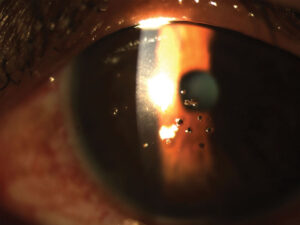
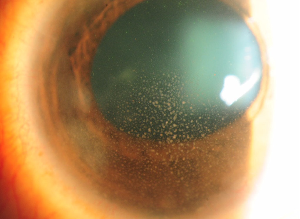
Mutton-Fat Keratic Precipitates
Mutton-fat KP are large, greasy, yellow-white deposits on the corneal endothelium. They contain macrophages and epithelioid cells, so they look thick and waxy.
They usually cluster inferiorly in Arlt triangle, and older deposits may become pigmented or glassy (see keratic precipitates).
All-fit Slit-Lamp Adapter
Record and share exactly what you see at the slit lamp. One adapter fits any slit lamp or surgical microscope — and any smartphone.
From Choroida — the team behind this siteIris Nodules in Granulomatous Uveitis
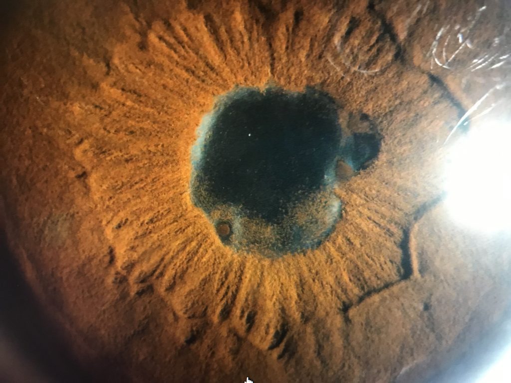
Koeppe nodules sit at the pupillary margin, while Busacca nodules lie on the anterior iris surface, away from the pupil.
Both represent small aggregates of inflammatory cells. Therefore, examine the iris carefully before dilating drops change the pupil margin.
Posterior Signs
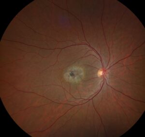
Granulomatous disease also involves the vitreous, retina and choroid, so look for snowballs, periphlebitis, choroidal granulomas and multifocal chorioretinal lesions.
Dalen-Fuchs nodules appear as small yellow spots deep to the retina (see Dalen-Fuchs nodules). As a result, every first visit needs a dilated fundus exam.
Why the Label Has Limits
Morphology overlaps between groups, and herpetic anterior uveitis, for example, can produce large KP.
Meanwhile, early topical steroids may shrink precipitates and blur the pattern, so interpret the label together with laterality, course and history.
Sarcoidosis in Granulomatous Uveitis
Sarcoidosis is among the most frequent causes, although frequency varies by region. Clues include bilateral disease, iris nodules, snowballs and candle-wax periphlebitis.
Also ask about cough, dyspnea, skin lesions and lacrimal gland swelling, while the IWOS criteria combine ocular signs with investigation results.
Tuberculosis
Tuberculosis mimics sarcoidosis closely, and broad posterior synechiae, choroidal granulomas and serpiginous-like choroiditis raise suspicion.
Then weigh endemic exposure, prior infection and immune status. Because treatment differs, avoid labeling sarcoid until TB is reasonably excluded.
Syphilis in Granulomatous Uveitis
Syphilis can produce almost any pattern of uveitis, and granulomatous anterior uveitis, placoid chorioretinitis and retinitis all occur.
Therefore, test every patient with granulomatous uveitis for syphilis, and also offer HIV testing, because coinfection changes management.
VKH and Sympathetic Ophthalmia
Both cause bilateral granulomatous panuveitis, often with serous retinal detachment. VKH occurs without trauma and may bring headache, tinnitus and later poliosis or vitiligo.
In contrast, sympathetic ophthalmia follows penetrating injury or intraocular surgery. For a side-by-side comparison, see VKH vs sympathetic ophthalmia.
Herpetic and Other Causes
Herpes simplex and varicella zoster uveitis may look granulomatous, especially when unilateral with raised pressure and sectoral iris atrophy. Next, consider lens-induced uveitis after capsule rupture or with a hypermature cataract. Finally, toxoplasmosis, fungal infection and multiple sclerosis-associated uveitis also belong on the list.
Baseline Tests for Granulomatous Uveitis
First, take a history covering trauma, eye surgery, travel, sexual history and TB exposure. Then order a core panel:
- Treponemal test plus RPR or VDRL titer
- Interferon-gamma release assay or tuberculin skin test
- Chest radiograph, with CT when suspicion persists
- Serum ACE and lysozyme, read with caution
- Serum calcium and liver enzymes when sarcoid is likely
Targeted Tests
Further tests depend on the clinical pattern. For example, aqueous PCR helps when herpetic or atypical infection is suspected. Biopsy of skin, conjunctival nodules or lymph nodes can confirm noncaseating granulomas.
Imaging in Granulomatous Uveitis
OCT detects macular edema, serous detachment and choroidal thickening, while indocyanine green angiography can reveal hypofluorescent choroidal dots missed on clinical exam.
B-scan ultrasound helps when media opacity hides the fundus, and imaging also provides a baseline for follow-up.
Treating Granulomatous Uveitis
Topical corticosteroids and cycloplegics control anterior inflammation and limit synechiae. However, steroids alone can worsen infection, so exclude syphilis and TB first whenever possible.
Posterior or bilateral disease often needs periocular, intravitreal or systemic corticosteroids, and chronic VKH and sarcoid frequently require steroid-sparing immunomodulatory therapy.
Counseling and Follow-Up in Granulomatous Uveitis
Explain that the term describes a pattern, while testing searches for the cause. Also warn that treatment may last months, with a slow taper to avoid rebound.
At each visit, monitor intraocular pressure, cataract, macular edema and new posterior lesions. When the response is unexpected, revisit the diagnosis.
Pitfalls to Avoid
- Do not start systemic steroids before syphilis and TB testing
- Do not rely on serum ACE alone to confirm or exclude sarcoidosis
- Never skip the dilated fundus exam in anterior disease
- Ask about recent eye surgery or trauma in bilateral cases


Document what you see
Two smartphone imaging tools built for everyday clinic use — one for the slit lamp, one for the fundus.
From Choroida — the team behind this siteReferences
- Jabs DA, Nussenblatt RB, Rosenbaum JT; Standardization of Uveitis Nomenclature (SUN) Working Group. Standardization of uveitis nomenclature for reporting clinical data. Am J Ophthalmol. 2005;140:509-516.
- Herbort CP, Rao NA, Mochizuki M. International criteria for the diagnosis of ocular sarcoidosis: results of the first International Workshop on Ocular Sarcoidosis (IWOS). Ocul Immunol Inflamm. 2009;17:160-169.
- Read RW, Holland GN, Rao NA, et al. Revised diagnostic criteria for Vogt-Koyanagi-Harada disease: report of an international committee on nomenclature. Am J Ophthalmol. 2001;131:647-652.
- Gupta V, Gupta A, Rao NA. Intraocular tuberculosis: an update. Surv Ophthalmol. 2007;52:561-587.
- American Academy of Ophthalmology. Basic and Clinical Science Course, Section 9: Uveitis and Ocular Inflammation. San Francisco: AAO; 2023.

Writes and reviews the clinical articles on Educate. Every post is checked against peer-reviewed sources before it goes live.