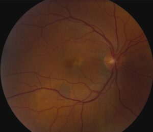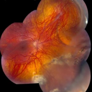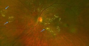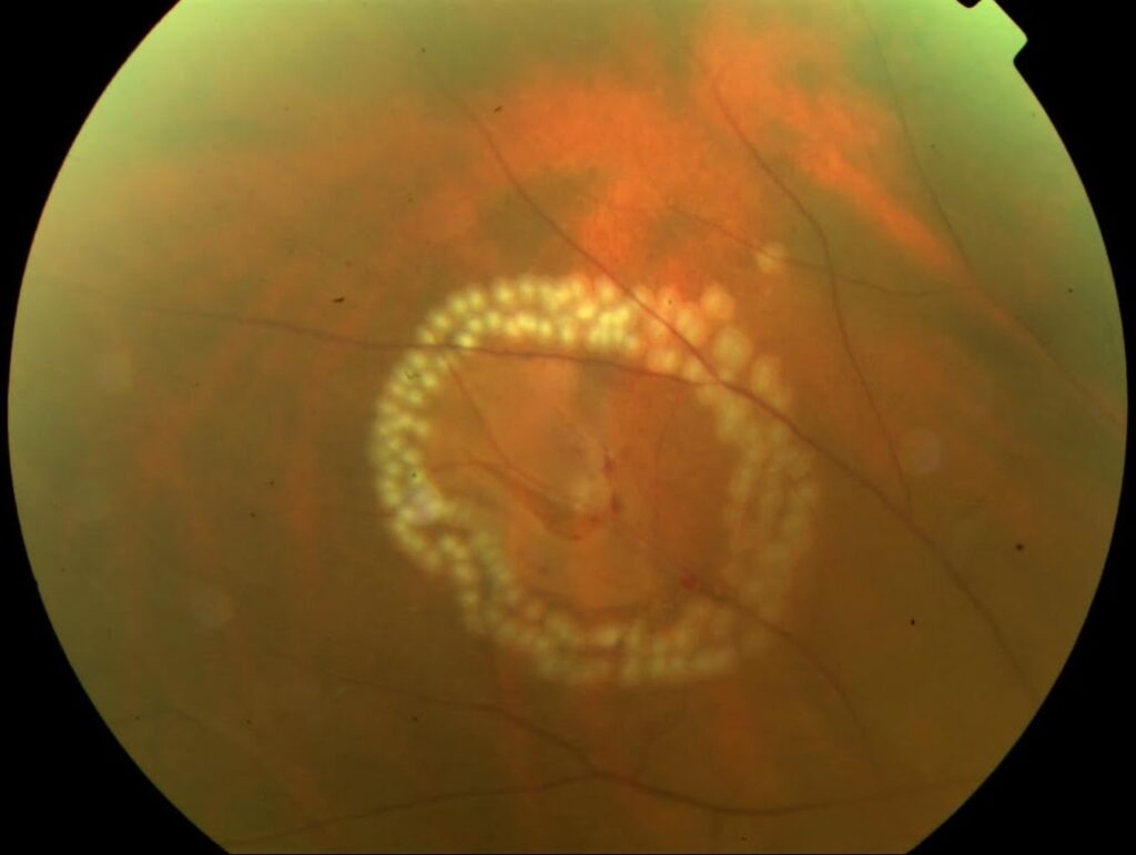
Hole vs tear is the first question to answer when you find a peripheral retinal break.
Both are full-thickness defects in the neurosensory retina.
However, they differ in mechanism, traction and detachment risk.
Therefore, break morphology and symptoms largely decide who needs laser.
Hole vs Tear: The Basic Difference
A hole usually reflects retinal thinning or a completed traction event.
A tear forms when vitreous traction rips the retina, and that pull often persists on a flap.
As a result, ongoing traction drives most of the risk.
Atrophic Holes
Atrophic holes are small, round breaks that arise from chronic retinal thinning.
They often sit inside lattice degeneration (see lattice with holes).
Most patients have no symptoms, so the finding usually appears at routine dilated examination.
However, young myopes need closer attention, because shallow detachments can develop slowly.
Fundus Explorer Pro
Photograph the retinal findings described here with the phone already in your pocket — 22 D optics and built-in illumination in one handheld unit.
From Choroida — the team behind this siteOperculated Holes
An operculated hole forms when vitreous traction pulls a plug of retina free.
Because traction has released, the risk of progression is generally low.
Still, inspect the edges, since residual vitreous attachment changes the assessment.
Flap Tears in Hole vs Tear
Flap (horseshoe) tears usually develop during posterior vitreous detachment, at sites of firm adhesion.
The flap stays hinged anteriorly while the vitreous keeps pulling on it.
Older natural history series suggest that about a third to a half of untreated symptomatic tears progress to detachment.
Hole vs Tear by Symptoms
New flashes and floaters suggest acute vitreous traction.
Therefore, search for a new break when a patient with a known hole reports them.
A shower of floaters may reflect vitreous hemorrhage from a torn vessel.
A curtain or field defect suggests that detachment has begun.
Hole vs Tear on Examination
First, check the anterior vitreous for pigment cells (Shafer sign), which strongly suggest a break.
Next, perform dilated indirect ophthalmoscopy with scleral depression through the full periphery.
In addition, look for several breaks, because one finding should never end the search.
Finally, examine the fellow eye.
Imaging in Hole vs Tear
Widefield photography documents the location and size of a peripheral break.
B-scan ultrasonography helps when hemorrhage blocks the view.
However, a normal image does not replace a careful depressed examination.
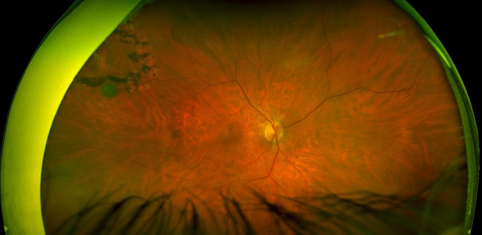
Which Breaks Need Laser
The AAO Preferred Practice Pattern advises prompt treatment for acute symptomatic flap tears.
By contrast, symptomatic operculated holes may not need treatment when traction has clearly released.
Asymptomatic atrophic and operculated holes rarely need treatment.
Asymptomatic flap tears can often be observed, although clinicians individualize that decision.
What Shifts the Hole vs Tear Decision
Several findings lower the threshold for treatment, even with a seemingly benign break.
For example, clinicians weigh subretinal fluid, high myopia, pseudophakia, and a detachment in the fellow eye.
However, evidence for prophylaxis is weak, and a Cochrane review found no adequate trials for asymptomatic breaks.
When Fluid Changes the Plan
A small cuff of subretinal fluid may suit surrounding laser.
Spreading fluid calls for retinal surgical review.
Demarcation lines suggest a chronic, slowly progressive process.
Also, recheck the macula, because symptoms can lag behind the anatomy.
What Laser Retinopexy Can and Cannot Do
Laser creates a chorioretinal adhesion around the break that matures over days (see laser retinopexy).
Cryotherapy offers an alternative when media opacity or an anterior location limits laser.
Laser reduces detachment risk, although the risk does not reach zero.
It does not remove vitreous traction, and it cannot prevent new breaks elsewhere.
Risks include new tears and epiretinal membrane.
Counseling for Hole vs Tear
Patients often hear “hole” and assume the worst.
Therefore, explain that many holes stay stable, while a fresh symptomatic tear needs quicker action.
Tell treated patients that floaters may persist after laser.
Give clear return instructions for new floaters, more flashes, a shadow or reduced vision.
Follow-Up After Hole vs Tear Assessment
A treated flap tear generally needs review within one to two weeks, then again over the following months.
A symptomatic PVD also deserves repeat examination when the first view was limited, because delayed breaks occur.
At each visit, depress the periphery again and check the laser scars for new fluid.
Pitfalls to Avoid
- Do not label a break atrophic without inspecting it for a flap
- Never dismiss Shafer sign after a limited peripheral view
- Do not attribute new symptoms to an old hole
- Avoid stopping the search after finding one break
- Do not promise that laser eliminates detachment risk


Document what you see
Two smartphone imaging tools built for everyday clinic use — one for the slit lamp, one for the fundus.
From Choroida — the team behind this siteReferences
- Flaxel CJ, Adelman RA, Bailey ST, et al. Posterior Vitreous Detachment, Retinal Breaks, and Lattice Degeneration Preferred Practice Pattern. Ophthalmology. 2020;127:P146-P181.
- Byer NE. What happens to untreated asymptomatic retinal breaks, and are they affected by posterior vitreous detachment? Ophthalmology. 1998;105:1045-1049.
- Davis MD. Natural history of retinal breaks without detachment. Arch Ophthalmol. 1974;92:183-194.
- Wilkinson CP. Interventions for asymptomatic retinal breaks and lattice degeneration for preventing retinal detachment. Cochrane Database Syst Rev. 2014;(9):CD003170.
- Coffee RE, Westfall AC, Davis GH, et al. Symptomatic posterior vitreous detachment and the incidence of delayed retinal breaks: case series and meta-analysis. Am J Ophthalmol. 2007;144:409-413.

Writes and reviews the clinical articles on Educate. Every post is checked against peer-reviewed sources before it goes live.