Metastasis vs melanoma is a common question when a clinician finds a solid choroidal mass in an adult.
Uveal metastasis is the commonest intraocular malignancy in adults, while melanoma is the commonest primary one. Both can elevate the retina and cause subretinal fluid. However, their shape, color, multiplicity and imaging behavior usually differ.
Metastasis vs Melanoma: Why the Label Changes Care
The diagnosis decides who manages the patient next, and melanoma needs ocular oncology treatment and lifelong surveillance for liver spread. In contrast, a metastasis signals disseminated cancer, so the oncologist leads, and a wrong label can therefore delay staging.
Start With the History
Ask about any previous cancer, even one treated decades ago, because breast carcinoma in women and lung carcinoma in men cause most choroidal metastases. Also ask about smoking, cough and weight loss, although many patients have no known primary when the eye lesion appears.
Fundus Explorer Pro
Photograph the retinal findings described here with the phone already in your pocket — 22 D optics and built-in illumination in one handheld unit.
From Choroida — the team behind this siteSymptoms Patients Report
Patients with metastasis often describe blurring that progresses over weeks, whereas melanoma, by comparison, is frequently asymptomatic and found on routine examination, so symptoms alone never settle the diagnosis.
Tumor Shape in Metastasis vs Melanoma
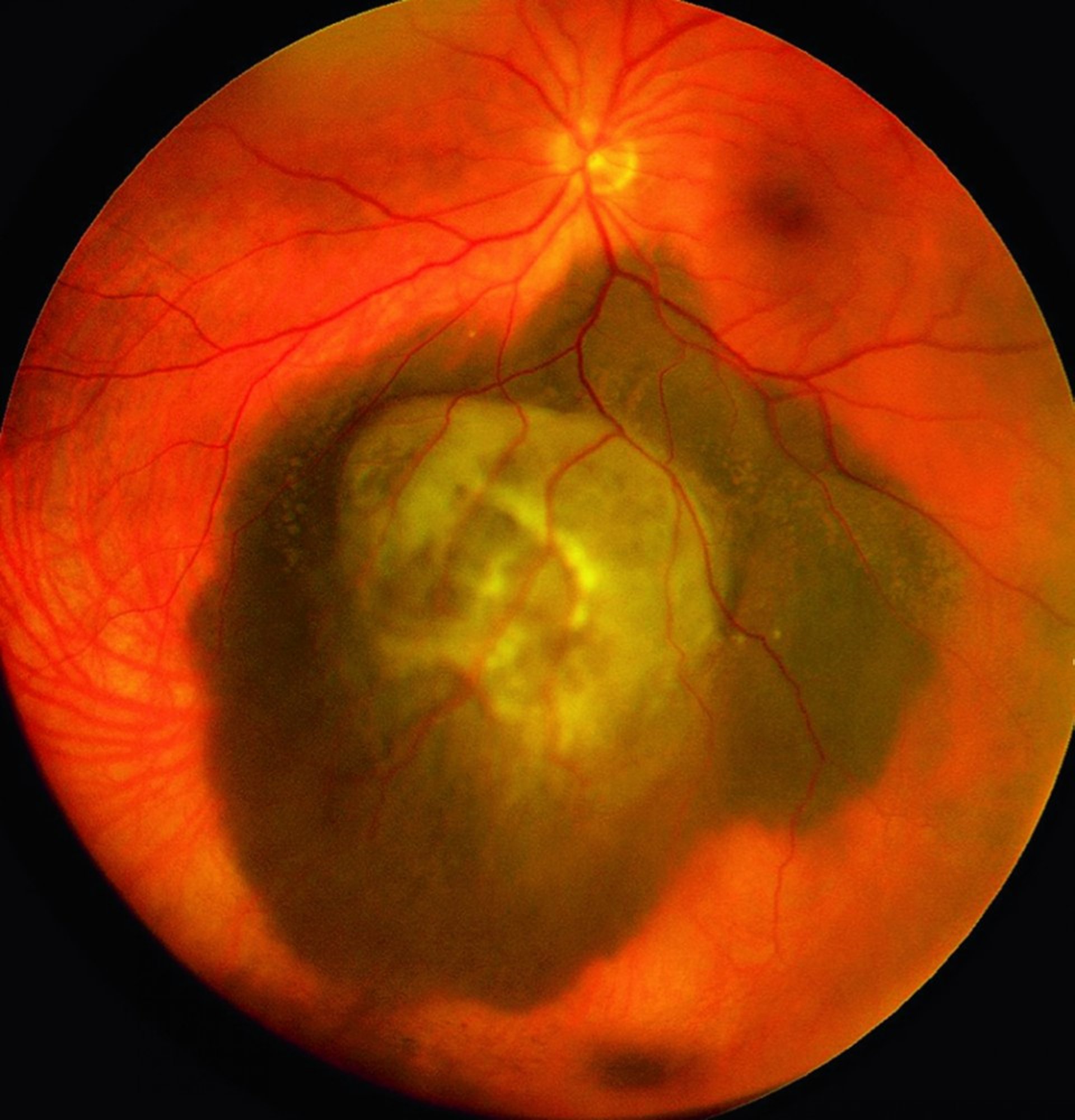
Metastases are typically placoid, with a flat or gently elevated profile and blurred margins, while melanoma usually forms a well-defined dome. After it breaks Bruch membrane, it can take a mushroom shape (see collar button melanoma), and that configuration strongly favors melanoma.
Color and Surface Clues
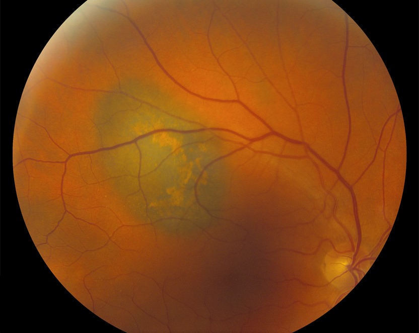
Most metastases look creamy yellow, and brown pigment clumps on the surface can also create a leopard spot pattern. However, carcinoid, renal and thyroid metastases often appear orange or red, while melanoma is usually brown or gray, although amelanotic tumors exist, and overlying orange pigment and drusen point toward a melanocytic lesion.
Multiplicity and Bilaterality
Multifocal or bilateral choroidal tumors strongly suggest metastasis, whereas choroidal melanoma is almost always unifocal and unilateral, so examine the fellow eye fully dilated.
Subretinal Fluid in Metastasis vs Melanoma
Metastases often produce extensive subretinal fluid for their thickness, and the fluid may shift with posture and detach the inferior retina. Melanoma also causes fluid, yet it usually stays closer to the tumor. Therefore, fluid out of proportion to a thin mass raises suspicion (see subretinal fluid on OCT).
B-Scan Ultrasound in Metastasis vs Melanoma
Melanoma typically shows acoustic hollowness and sometimes choroidal excavation, while a metastasis usually lacks hollowness and has an irregular, lumpy surface, although both may show an exudative retinal detachment.
A-Scan Reflectivity in Metastasis vs Melanoma
Melanoma displays low to medium internal reflectivity with a regular structure, while metastases more often show medium to high, irregular reflectivity. However, values overlap, so interpret them with the clinical picture.
OCT and EDI-OCT Signs
Enhanced depth imaging displays the anterior tumor contour, and metastases commonly show a lumpy bumpy anterior surface. Small melanomas more often have a smooth, dome-shaped contour, although both may show shaggy photoreceptors, so OCT supports the diagnosis without proving it.
Angiography in Metastasis vs Melanoma
Many metastases show early hypofluorescence and late diffuse staining on fluorescein angiography. Large melanomas, by comparison, may show a double circulation from intrinsic tumor vessels, while on indocyanine green angiography, metastases tend to stay hypofluorescent.
Growth Rate and Follow-Up
Metastases can enlarge noticeably within weeks, while melanoma usually grows more slowly. Therefore, short-interval photography and ultrasound help when the first visit is equivocal.
When to Biopsy
Fine needle aspiration biopsy helps when imaging and history remain inconclusive, for example because immunohistochemistry may suggest breast or lung origin, while melanoma tissue allows gene expression profiling for prognosis.
Systemic Workup
A suspected metastasis needs prompt oncology referral and body imaging, often PET-CT. Melanoma also needs staging, mainly liver imaging (see choroidal melanoma), so both teams can coordinate ocular and systemic care.
How Treatment Differs
Metastases often respond to systemic chemotherapy, targeted therapy or immunotherapy, and external beam radiotherapy, plaque radiotherapy or photodynamic therapy can control symptomatic lesions. Melanoma, by contrast, needs plaque brachytherapy, proton beam therapy or enucleation. Explain to the patient that ocular treatment protects vision and comfort, while survival depends on systemic disease.
Pitfalls to Avoid
- Do not skip a cancer history in an older patient with a yellow mass
- Do not assume every amelanotic dome is a metastasis
- Never overlook lesions in the fellow eye
- Do not rely on a single imaging test
- Avoid delaying referral while waiting for growth in a symptomatic eye


Document what you see
Two smartphone imaging tools built for everyday clinic use — one for the slit lamp, one for the fundus.
From Choroida — the team behind this siteReferences
- Shields CL, Shields JA, Gross NE, Schwartz GP, Lally SE. Survey of 520 eyes with uveal metastases. Ophthalmology. 1997;104:1265-1276.
- Shields CL, Welch RJ, Malik K, et al. Uveal metastasis: clinical features and survival outcome of 2214 tumors in 1111 patients based on primary tumor origin. Middle East Afr J Ophthalmol. 2018;25:81-90.
- Al-Dahmash SA, Shields CL, Kaliki S, Johnson T, Shields JA. Enhanced depth imaging optical coherence tomography of choroidal metastasis in 14 eyes. Retina. 2014;34:1588-1593.
- Collaborative Ocular Melanoma Study Group. Accuracy of diagnosis of choroidal melanomas in the Collaborative Ocular Melanoma Study. COMS report no. 1. Arch Ophthalmol. 1990;108:1268-1273.
- Shields CL, Kaliki S, Rojanaporn D, et al. Enhanced depth imaging optical coherence tomography of small choroidal melanoma: comparison with choroidal nevus. Arch Ophthalmol. 2012;130:850-856.
Test yourself
A few questions straight from this article.
-
What is the commonest intraocular malignancy in adults?
Uveal metastasis is the commonest intraocular malignancy in adults, while melanoma is the commonest primary one. -
Which primary cancers cause most choroidal metastases?
Breast carcinoma in women and lung carcinoma in men cause most choroidal metastases, so ask about any previous cancer, even one treated decades ago. -
What tumour profile is typical of a choroidal metastasis?
Metastases are typically placoid with a flat or gently elevated profile and blurred margins, while melanoma usually forms a well-defined dome. -
Finding bilateral or multifocal choroidal tumours most strongly suggests which diagnosis?
Multifocal or bilateral tumours strongly suggest metastasis, whereas choroidal melanoma is almost always unifocal and unilateral, so the fellow eye must be examined dilated. -
Which subretinal fluid pattern raises suspicion of choroidal metastasis rather than melanoma?
Metastases often produce extensive, shifting subretinal fluid for their thickness, while melanoma fluid usually stays closer to the tumour. -
What anterior tumour contour does enhanced depth imaging OCT commonly show in choroidal metastasis?
EDI-OCT commonly shows a lumpy bumpy anterior surface in metastases, whereas small melanomas more often have a smooth dome-shaped contour. -
How do choroidal metastases tend to appear on indocyanine green angiography?
On ICG angiography metastases tend to stay hypofluorescent; on FA many show early hypofluorescence and late diffuse staining, while large melanomas may show a double circulation. -
How does the growth rate of choroidal metastasis compare with melanoma?
Metastases can enlarge within weeks while melanoma usually grows more slowly, so short-interval photography and ultrasound help when the first visit is equivocal. -
When imaging is inconclusive, what can fine needle aspiration biopsy of a melanoma provide?
Biopsy helps when history and imaging are inconclusive: immunohistochemistry may suggest breast or lung origin, and melanoma tissue allows gene expression profiling for prognosis. -
Staging for choroidal melanoma mainly relies on imaging of which organ?
A suspected metastasis needs oncology referral and body imaging, often PET-CT, while melanoma staging is mainly liver imaging, reflecting its lifelong risk of liver spread.

Writes and reviews the clinical articles on Educate. Every post is checked against peer-reviewed sources before it goes live.