Multicolor imaging is a confocal scanning laser ophthalmoscopy technique that captures the fundus with three laser wavelengths at once.
The device then merges the three reflectance images into one composite picture. The result resembles a color photo, yet it is a pseudocolor image built from separate depth signals, so good reading starts with knowing what each channel contributes.
How Multicolor Imaging Works
The Spectralis platform scans the retina with blue (488 nm), green (515 nm) and infrared (820 nm) lasers simultaneously, while a confocal pinhole rejects light scattered from outside the focal plane. As a result, the image keeps high contrast and suffers less blur from media haze, and software then assigns each reflectance image to a color channel and combines them.
Why Wavelength Sets Depth
Light penetrates the fundus more deeply as wavelength increases, and blue light scatters in the inner retina, so it reports mostly superficial structure. Green light reaches slightly deeper, while infrared light reflects from the outer retina, RPE and choroid.
Fundus Explorer Pro
Photograph the retinal findings described here with the phone already in your pocket — 22 D optics and built-in illumination in one handheld unit.
From Choroida — the team behind this siteThe Blue Channel in Multicolor Imaging
Blue reflectance highlights the nerve fiber layer and the internal limiting membrane surface, while macular pigment absorbs blue light, so the fovea looks dark. Therefore, this channel suits epiretinal membranes, macular holes and nerve fiber layer defects.
The Green Channel
Hemoglobin absorbs green light strongly, so retinal vessels, hemorrhages and microaneurysms show sharp contrast, and hard exudates also stand out, which helps in diabetic retinopathy.
The Infrared Channel
Infrared reflectance reveals drusen, pigment changes and subretinal fluid, and it also tolerates cataract and small pupils better than visible light.
Reading the Composite Multicolor Image
The merged image usually shows a greenish tone over a reddish background, and superficial lesions tend to look green or cyan, while deeper lesions look orange or red. However, these colors differ from true fundus colors, so blood and pigment may look unfamiliar. Therefore, open the individual channels before you interpret any color change.
Epiretinal Membrane on Multicolor Imaging
An epiretinal membrane appears as a bright green sheen with radiating retinal folds, and the blue channel shows the membrane edge and striae clearly. Muftuoglu and colleagues reported better visualization of macular pucker with multicolor imaging than with color photography (see epiretinal membrane), although OCT still defines thickness and traction.
Reticular Pseudodrusen in Multicolor Imaging
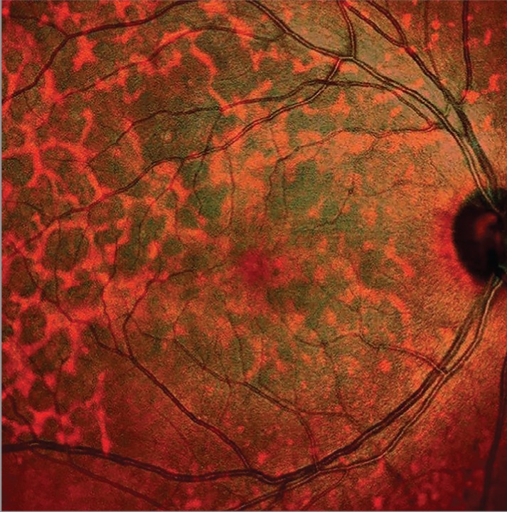
Reticular pseudodrusen are often subtle on color photographs, whereas multicolor imaging can show a regular network of round lesions, often with a target aspect. Infrared reflectance supplies most of this contrast (see reticular pseudodrusen).
Subretinal Fluid and Central Serous Chorioretinopathy
In central serous chorioretinopathy, a serous detachment often appears as a well-demarcated area of altered color, so multicolor imaging can outline the extent of fluid and help track resolution. However, it cannot measure fluid height or show leakage, so OCT and angiography remain necessary (see subretinal fluid on OCT).
Geographic Atrophy and AMD
Atrophic areas show sharp borders on the composite image, and Ben Moussa and colleagues evaluated multicolor imaging for geographic atrophy and compared it with autofluorescence. In daily practice, autofluorescence remains the usual method for following atrophy growth (see fundus autofluorescence).
Multicolor Imaging Versus Color Fundus Photography
Color photography uses white flash light and records true color from all layers together, while multicolor imaging separates depth information and offers higher contrast for some lesions. However, grading scales such as diabetic retinopathy severity rely on true color photos, and clinicians also judge pigment, lipid and disc pallor more reliably on true color (see fundus photography).
Artifacts in Multicolor Imaging
- Ghost maculopathy, a reflection artifact that can mimic a macular lesion
- Color imbalance from poor focus on the plane of interest
- Motion artifacts and line shifts during scanning
- A strong green sheen from a normal young internal limiting membrane
- Darkening toward the edges of the field
Practical Acquisition Tips
First, focus carefully on the layer you want to study, and next, average several frames to reduce noise. Also, keep the head straight, because tilt changes reflectance across the field.
Where Multicolor Imaging Fits
The same platform acquires OCT, so the composite image helps register scans to visible landmarks and also documents lesions well over time. However, it does not replace angiography, autofluorescence or true color photography.
Counseling the Patient
Explain that the scan uses low-power lasers and takes seconds, although patients may notice bright blue and green flashes. Then reassure them that the green tint is artificial and does not signal disease.
Pitfalls to Avoid
- Do not diagnose disease from composite color alone
- Do not apply photo-based grading scales to multicolor images
- Check each channel before calling a lesion superficial or deep
- Recognize ghost maculopathy before ordering further tests
- Confirm fluid and membranes with OCT


Document what you see
Two smartphone imaging tools built for everyday clinic use — one for the slit lamp, one for the fundus.
From Choroida — the team behind this siteReferences
- Tan ACS, Fleckenstein M, Schmitz-Valckenberg S, Holz FG. Clinical application of multicolor imaging technology. Ophthalmologica. 2016;236:8-18.
- Kilic Muftuoglu I, Bartsch DU, Barteselli G, et al. Visualization of macular pucker by multicolor scanning laser imaging. Retina. 2018;38:352-358.
- Alten F, Clemens CR, Heiduschka P, Eter N. Characterisation of reticular pseudodrusen and their central target aspect in multi-spectral, confocal scanning laser ophthalmoscopy. Graefes Arch Clin Exp Ophthalmol. 2014;252:715-721.
- Ben Moussa N, Georges A, Capuano V, et al. MultiColor imaging in the evaluation of geographic atrophy due to age-related macular degeneration. Br J Ophthalmol. 2015;99:842-847.
- Pang CE, Freund KB. Ghost maculopathy: an artifact on near-infrared reflectance and multicolor imaging masquerading as chorioretinal pathology. Am J Ophthalmol. 2014;158:171-178.
Test yourself
A few questions straight from this article.
-
Which three laser wavelengths does multicolor imaging use to build its composite fundus image?
The Spectralis platform scans with blue (488 nm), green (515 nm) and infrared (820 nm) lasers simultaneously, and software merges the reflectance images into one composite. -
Why is a multicolor imaging composite considered a pseudocolor image rather than a true color photograph?
Multicolor imaging merges three separate reflectance images into a composite that resembles a color photo but is built from separate depth signals. -
In multicolor imaging, which laser channel reflects mainly from the outer retina, RPE and choroid?
Light penetrates deeper as wavelength increases. Blue reports superficial structure, green reaches slightly deeper, and infrared reflects from the outer retina, RPE and choroid. -
Why does the fovea look dark on the blue channel of multicolor imaging?
Blue reflectance highlights the nerve fiber layer and ILM surface, while macular pigment absorbs blue light, so the fovea appears dark. -
In multicolor imaging, which channel shows retinal vessels, hemorrhages and microaneurysms with sharp contrast in diabetic retinopathy?
Hemoglobin absorbs green light strongly, so vessels, hemorrhages and microaneurysms show sharp contrast, and hard exudates also stand out. -
On a multicolor imaging composite, how do superficial lesions typically look compared with deeper ones?
Superficial lesions tend to look green or cyan, while deeper lesions look orange or red. Individual channels should be checked before interpreting color changes. -
How does an epiretinal membrane typically appear on multicolor imaging?
An epiretinal membrane appears as a bright green sheen with radiating retinal folds, and the blue channel shows the membrane edge and striae. OCT still defines thickness and traction. -
Which multicolor imaging finding suggests reticular pseudodrusen in age-related macular degeneration?
Reticular pseudodrusen are subtle on color photographs, but multicolor imaging can show a regular network of round lesions, often target-like, with infrared supplying most of the contrast. -
What is a limitation of multicolor imaging in central serous chorioretinopathy?
Multicolor imaging can outline fluid extent and help track resolution, but it cannot measure fluid height or show leakage, so OCT and angiography remain necessary. -
Which imaging method remains the usual choice for following geographic atrophy growth, rather than multicolor imaging?
Although atrophic areas show sharp borders on multicolor imaging, autofluorescence remains the usual method for following atrophy growth in daily practice.

Writes and reviews the clinical articles on Educate. Every post is checked against peer-reviewed sources before it goes live.