CASE REPORT
Case of Optociliary Shunt Vessels a 46-year-old man complained of severe visual loss starting 2 years earlier, originally without headache or eye pain. The first manifestation was transient visual obscuration. This was followed a few months later by the loss of the peripheral field, but with preservation of central vision.
Six months before the first examination, severe headache set in, associated with reduced central vision. The patient’s body mass index (BMI) was 26.5 kg/m2.
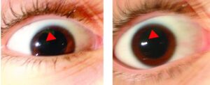
Upon examination, visual acuity was no light perception in the right eye and hand movements in the temporal field of the left eye. The pupillary light reflex was poor in both eyes. No abnormality was seen on biomicroscopy, and the intraocular pressure was 12 mm Hg in both eyes.
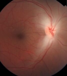
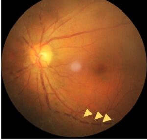
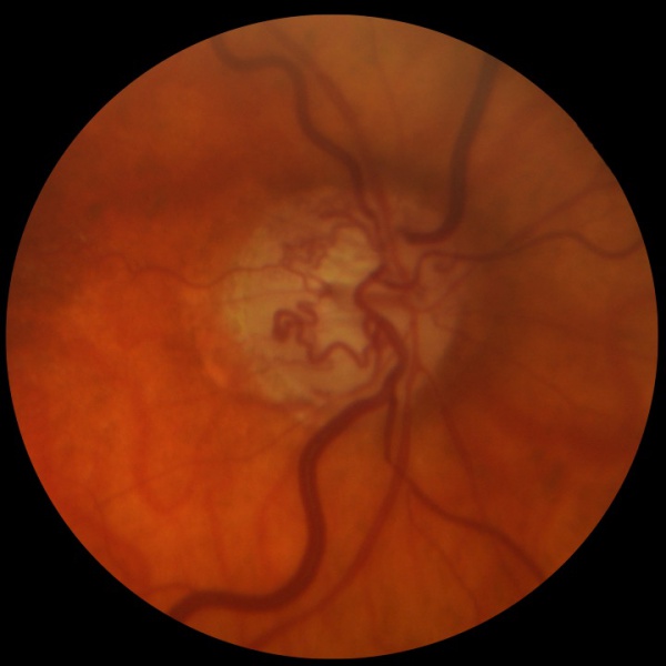
The fundus examination revealed atrophic optic nerve and optociliary shunt vessels in both eyes.
DISEASE of Optociliary Shunt Vessels
This disease also known as retinochoroidal shunt vessels of the optic disc or retinochoroidal collaterals, are collateral vessels on the optic nerve that connect the choroidal and retinal circulations.
They are associated with multiple conditions including central retinal vein occlusion, optic nerve sheath meningioma, chronic glaucoma, and chronic papilledema.

Fundus Explorer Pro
Photograph the retinal findings described here with the phone already in your pocket — 22 D optics and built-in illumination in one handheld unit.
From Choroida — the team behind this siteMANAGEMENT of Optociliary Shunt Vessels
Work-up and treatment are directed towards the underlying etiology. Optociliary shunt vessels classically do not disappear. However, various case reports have demonstrated shunt vessels to decrease in caliber or even completely involute after treatments that improve blood flow through the central retinal vein.
Another possibility is that the development of optic atrophy results in a thinner nerve with less obstruction, allowing a decrease in the central retinal vein pressure and lessening the driving force for blood flow through the optociliary collateral vessels.
The regression of optociliary shunt vessels has been documented in cases of pseudotumor cerebri successfully treated with optic nerve sheath fenestration.
Fundus photography is superior to fundus analysis as it enables intraocular pathologies to be photo captured and encrypted information to be shared with colleagues and patients.
Recent technologies allow smartphone-based attachments and integrated lens adaptors to transform the smartphone into a portable fundus camera.
More report cases of Aicardi syndrome, Choroidal Folds, Choroidal Osteoma, Cornea Verticillate.


Document what you see
Two smartphone imaging tools built for everyday clinic use — one for the slit lamp, one for the fundus.
From Choroida — the team behind this siteREFERENCES
- Fraser CL, Ridha MA, Biousse V, Newman NJ. Vitreous hemorrhage secondary to optociliary shunt vessels from papilledema. J Neuroophthalmol. Dec 2012;32(4):332-4. doi:10.1097/WNO.0b013e31825ba161
- Lee JJ, Yap EY. Optociliary shunt vessels in diabetes mellitus. Singapore Med J. Apr 2004;45(4):166-9.
- Takahashi K, Muraoka K, Kishi S, Shimizu K. Formation of retinochoroidal collaterals in central retinal vein occlusion. Am J Ophthalmol. Jul 1998;126(1):91-9. doi:10.1016/s0002-9394(98)00069-5.
- Muci-Mendoza R, Arevalo JF, Ramella M, et al. Optociliary veins in optic nerve sheath meningioma. Indocyanine green videoangiography findings. Ophthalmology. Feb 1999;106(2):311-8. doi:10.1016/s0161-6420(99)90055-6.
- de Alba Campomanes AG, Larson DA, Horton JC. Immediate shrinkage of optociliary shunt vessels after fractionated external beam radiation for meningioma of the optic nerve sheath. AJNR Am J Neuroradiol. Aug 2008;29(7):1360-2. doi:10.3174/ajnr.A1063.
- Interlandi E, Pellegrini F, Papayannis A, et al. Optical Coherence Tomography Angiography Findings in Optic Nerve Sheath Meningioma. Case Rep Ophthalmol. 2020;11(2):364-369. Published 2020 Jul 14. doi:10.1159/000508411
- Singh A, Agarwal A, Mahajan S, Karkhur S, Singh R, Bansal R, Dogra MR, Gupta V. Morphological differences between optic disc collaterals and neovascularization on optical coherence tomography angiography. Graefes Arch Clin Exp Ophthalmol. 2017 Apr;255(4):753-759. doi: 10.1007/s00417-016-3565-x. Epub 2016 Dec 9. PMID: 27942950.
Test yourself
A few questions straight from this article.
-
Optociliary shunt vessels connect which two circulations at the optic nerve?
Optociliary shunt vessels are collateral vessels on the optic nerve that connect the choroidal circulation to the retinal circulation. -
Which alternative names are used for optociliary shunt vessels?
The article notes the condition is also called retinochoroidal shunt vessels of the optic disc, or retinochoroidal collaterals. -
Which condition is listed as an association of optociliary shunt vessels?
Associations include central retinal vein occlusion, optic nerve sheath meningioma, chronic glaucoma and chronic papilledema. -
In the reported case of optociliary shunt vessels, what was the first visual symptom?
The 46-year-old man's first manifestation was transient visual obscuration, followed months later by peripheral field loss with preserved central vision. -
What was the recorded intraocular pressure in both eyes in this optociliary shunt vessel case?
Biomicroscopy showed no abnormality and the intraocular pressure was 12 mm Hg in both eyes. -
What did fundus examination reveal in this patient with optociliary shunt vessels?
Fundus examination revealed an atrophic optic nerve together with optociliary shunt vessels in both eyes. -
What is the recorded visual acuity of the right eye in this optociliary shunt vessel case?
Visual acuity was no light perception in the right eye and hand movements in the temporal field of the left eye, with poor pupillary light reflexes bilaterally. -
How should a patient with optociliary shunt vessels be managed?
Work-up and treatment are directed towards the underlying etiology rather than at the shunt vessels themselves. -
What is the classic natural history of optociliary shunt vessels once formed?
Optociliary shunt vessels classically do not disappear, although case reports describe reduction in calibre or involution after treatments that improve central retinal vein flow. -
Regression of optociliary shunt vessels has been documented after which procedure for pseudotumor cerebri?
The article documents regression of optociliary shunt vessels in cases of pseudotumor cerebri successfully treated with optic nerve sheath fenestration. -
How may optic atrophy explain reduced flow through optociliary shunt vessels?
Optic atrophy produces a thinner nerve with less obstruction, decreasing central retinal vein pressure and lessening the driving force for flow through the collaterals.

Writes and reviews the clinical articles on Educate. Every post is checked against peer-reviewed sources before it goes live.