
Case report
A 28-year-old woman referred to a clinic with complaints of decreased vision without night blindness and visual acuity of both eyes was 20/25 with plano refraction.
Her slit-lamp examination revealed clear cornea and clear lens, and 1+ pigment in vitreous. Intraocular pressure was 18 mmHg for the right eye and 17 mmHg for the left eye. She did not have any history of associated other medical conditions or drug usage.
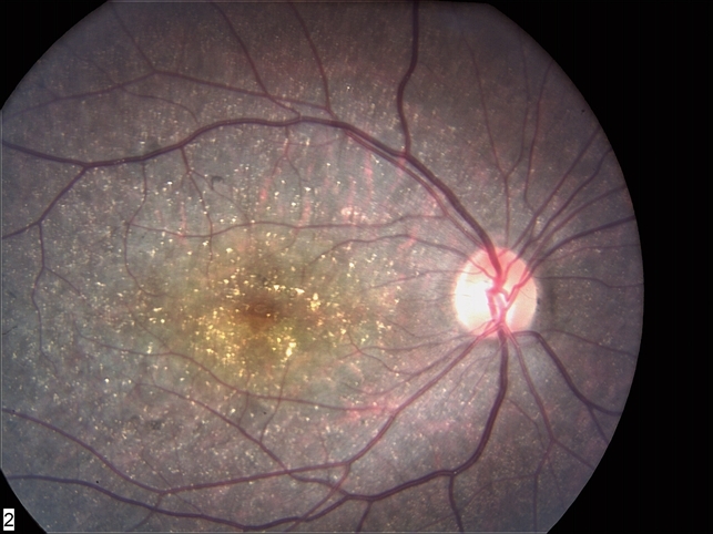
Fundus examination showed intraretinal crystals distributed in the posterior pole and also mid periphery. There was mid-peripheral RPE and choriocapillaris atrophy, peripheral pigment clumping, and retinal scarring.
Fluorescein angiography highlighted the focal geographic appearance showing transmission hyperfluorescence in the crystalline retina and choriocapillaris atrophy in the adjacent noncrystalline retina. A diagnosis of Bietti Crystalline Dystrophy was made.
Disease
Bietti’s Crystalline Dystrophy (BCD), is a rare autosomal recessive ocular disease that involves yellow-white crystalline lipid deposits in the retina and sometimes cornea, degeneration of the retinal pigment epithelium (RPE), and sclerosis of the choroidal vessels.
Progression of the disease ultimately results in reduced visual acuity, night blindness, visual field loss, and impaired color vision. The onset of the disease can occur from the early teenage years to the third decade of life, but can also occur beyond the third decade.
As the disease progresses, decreases in peripheral acuity, central acuity, or both ultimately result in legal blindness in most patients.
Fundus Explorer Pro
Photograph the retinal findings described here with the phone already in your pocket — 22 D optics and built-in illumination in one handheld unit.
From Choroida — the team behind this siteClinical presentation
Typically patients with BCD begin noticing symptoms between the second and third decades of life. Patients experience a progressive reduction in visual acuity, nyctalopia, and visual field loss.
Occasionally, color vision may also be impaired. Visual deficits can progress at different rates in each eye, and the severity and rate of progression vary widely among affected individuals.

Conclusion
Bietti’s Crystalline Dystrophy (BCD) is rare retinal dystrophy that is characterized by the deposition of yellow-white crystals in the retina and sometimes the cornea, degeneration of the retina, and sclerosis of the choroidal vessels that results in the progressive loss of peripheral visual acuity and night blindness.
There is currently no specific treatment for BCD. However, early diagnosis and referral to low-vision specialists can help with the management of patients with the disease.
Fundus photography is superior to fundus analysis as it enables intraocular pathologies to be photo captured and encrypted information to be shared with colleagues and patients.
Recent technologies allow smartphone-based attachments and integrated lens adaptors to transform the smartphone into a portable fundus camera.
More about: Christmas tree cataract, Retinitis Pigmentosa, Branch Retinal Artery Occlusion, Coats disease.


Document what you see
Two smartphone imaging tools built for everyday clinic use — one for the slit lamp, one for the fundus.
From Choroida — the team behind this siteReferences
- American Academy of Ophthalmology. Bietti crystalline dystrophy. Accessed September 19, 2019
- Bietti G. Ueber familiaeres Vorkommen von ‘Retinitis punctata albescens’ (verbunden mit ‘Dystrophia marginalis cristallinea corneae’), Glitzern des Glaskoerpers und anderen degenerativen Augenveraenderungen. Klin Monatsbl Augenheilkd. 99: 737-757, 1937.
- Lin J, Nishiguchi KM, Nakamura M, Dryja TP, Berson EL, Miyake Y. Recessive mutations in the CYP4V2 gene in East Asian and Middle Eastern patients with Bietti crystalline corneoretinal dystrophy. J Med Genet. 2005;42(6):e38.
- Kaiser-Kupfer MI, Chan CC, Markello TC, Crawford MA, Caruso RC, Csaky KG, Guo J. Clinical biochemical and pathological correlations in Bietti’s crystalline dystrophy. Am J Ophthalmol 1994; 118: 569–582.
- Lee J, Jiao X, Hejtmancik JF, et al. The metabolism of fatty acids in human Bietti crystalline dystrophy. Invest Ophthalmol Vis Sci. 2001;42(8):1707-14.
Test yourself
A few questions straight from this article.
-
What is the inheritance pattern of Bietti crystalline dystrophy?
Bietti crystalline dystrophy is a rare autosomal recessive ocular disease, so parents are typically unaffected carriers. -
What kind of deposits characterise Bietti crystalline dystrophy?
The hallmark is yellow-white crystalline lipid deposits within the retina, which may also involve the cornea in some patients. -
Besides crystal deposition, which structures degenerate in Bietti crystalline dystrophy?
The disease combines crystal deposition with degeneration of the retinal pigment epithelium and sclerosis of the choroidal vessels. -
When do patients with Bietti crystalline dystrophy usually begin noticing symptoms?
Onset ranges from the early teenage years into the third decade, and occasionally later, with most patients becoming symptomatic in their twenties. -
How are the retinal crystals of Bietti crystalline dystrophy distributed on fundus examination?
Intraretinal crystals are seen scattered across both the posterior pole and the mid periphery of the fundus. -
What does fluorescein angiography show in Bietti crystalline dystrophy?
Angiography highlights a focal geographic pattern, with transmission hyperfluorescence over crystalline retina and choriocapillaris atrophy in adjacent non-crystalline retina. -
Which symptom cluster develops as Bietti crystalline dystrophy progresses?
Progression brings reduced visual acuity, nyctalopia and visual field loss, with colour vision occasionally impaired as well. -
What is the usual long-term visual outcome in Bietti crystalline dystrophy?
Loss of peripheral acuity, central acuity or both progresses until most affected patients meet criteria for legal blindness. -
What does management of Bietti crystalline dystrophy currently consist of?
No specific treatment exists for the disease, so early diagnosis and referral to low-vision specialists form the basis of management.

Writes and reviews the clinical articles on Educate. Every post is checked against peer-reviewed sources before it goes live.



