Carotid stenosis eye findings are often the first visible sign of atherosclerosis in the internal carotid artery.
The eye receives blood through the ophthalmic artery, which also links it to cerebral circulation. Therefore, a careful fundus examination can reveal stroke risk before any neurological event.
Why Carotid Stenosis Eye Findings Matter
The ophthalmic artery is the first major branch of the internal carotid artery. As a result, carotid plaque can harm the eye in two ways. First, fragments break off and lodge in retinal arterioles, and second, severe narrowing lowers perfusion pressure to the whole globe.
Retinal Emboli as a Carotid Stenosis Eye Sign
Hollenhorst plaques are bright, refractile, yellow cholesterol crystals at arteriolar bifurcations, and they usually come from ulcerated carotid atheroma. In contrast, dull white calcific emboli suggest a cardiac valve source. Even an asymptomatic plaque marks systemic atherosclerosis, so photograph it and arrange vascular evaluation (see retinal emboli types and workup).
Fundus Explorer Pro
Photograph the retinal findings described here with the phone already in your pocket — 22 D optics and built-in illumination in one handheld unit.
From Choroida — the team behind this siteAmaurosis Fugax in Carotid Stenosis Eye Disease
Patients describe painless monocular dimming, often like a curtain, lasting seconds to minutes, after which vision returns and the fundus may look normal. Because the episode reflects transient retinal ischemia, current definitions treat it as a transient ischemic attack. Also confirm that only one eye was affected, since a homonymous field defect points to the brain.
Retinal Artery Occlusion
Central or branch retinal artery occlusion is a retinal stroke, and the retina shows whitening, sometimes with a visible embolus or a cherry-red spot. In addition, these patients carry a raised short-term risk of cerebral stroke. Carotid plaque is a common source, while cardiac embolism and arteritis remain alternatives.
Venous Stasis Retinopathy
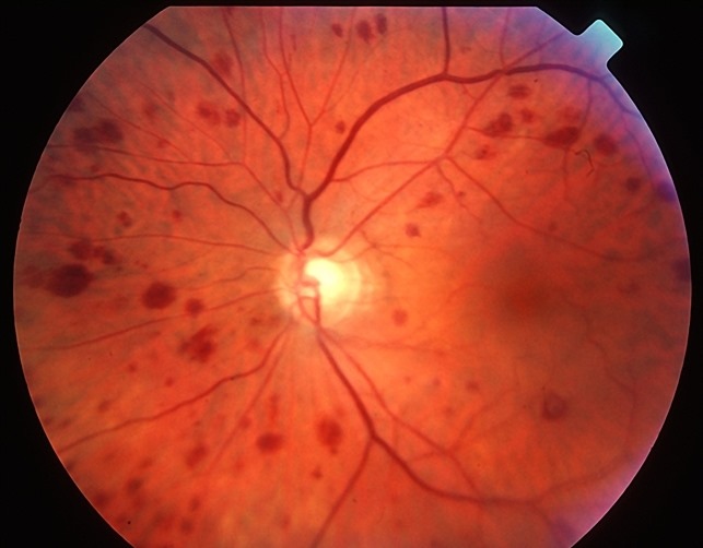
Moderate hypoperfusion produces dilated veins and scattered dot and blot hemorrhages in the midperiphery. The picture resembles a mild vein occlusion, although the disc usually looks normal. If perfusion falls further, it can progress to ocular ischemia (see venous stasis retinopathy).
Ocular Ischemic Syndrome as a Carotid Stenosis Eye Emergency
Ocular ischemic syndrome reflects chronic, severe hypoperfusion of the entire eye, and classic series describe carotid stenosis of 90 percent or more in most patients. Patients report gradual vision loss, slow recovery after bright light, and a dull orbital ache.
Signs include narrowed arteries, midperipheral hemorrhages, iris neovascularization and mild anterior chamber flare. Then pressure may run low from ciliary body ischemia, or high from neovascular glaucoma.
Telling Ischemia From Vein Occlusion
Both conditions show hemorrhages and dilated veins, although ischemic veins are rarely tortuous and hemorrhages favor the midperiphery. Light digital pressure on the globe often induces arterial pulsation in ischemia. Fluorescein angiography also shows delayed choroidal filling and prolonged arteriovenous transit (see OIS vs CRVO).
Asymmetric Retinopathy in Carotid Stenosis Eye Cases
Diabetic retinopathy that is clearly worse in one eye deserves a second look, while severe stenosis can make the ipsilateral eye more ischemic. Conversely, low perfusion pressure may spare that eye from hypertensive changes. Therefore, unexplained asymmetry justifies carotid imaging.
History and Risk Factors
Ask about prior strokes, transient weakness, speech disturbance or earlier episodes of monocular blur. Next, review vascular risk factors:
- Smoking
- Hypertension and diabetes
- Dyslipidemia
- Coronary or peripheral arterial disease
Excluding Giant Cell Arteritis
Patients over 50 with artery occlusion or transient vision loss need assessment for arteritis, so ask about headache, jaw claudication and scalp tenderness. Then order sedimentation rate, C-reactive protein and platelet count urgently, because untreated arteritis can blind the second eye within days.
Triage of Carotid Stenosis Eye Findings
Urgency depends on symptoms and timing.
- Acute artery occlusion or recent amaurosis fugax: same-day emergency stroke assessment
- Ocular ischemic syndrome: urgent carotid imaging and vascular surgery referral
- Venous stasis retinopathy: prompt imaging with close retinal review
- Asymptomatic emboli: outpatient vascular workup without long delay
Carotid Imaging and Workup
Carotid duplex ultrasound is the usual first test. CT or MR angiography then defines stenosis grade before intervention, and you should also request an ECG, echocardiography, lipid profile and glucose.
Treatment and Its Limits
NASCET showed that endarterectomy reduced stroke in symptomatic patients with 70 to 99 percent stenosis, and antiplatelet therapy, statins and risk factor control apply to almost everyone. However, revascularization may not reverse established ocular ischemia, and pressure can even rise after surgery because aqueous production recovers. Panretinal photocoagulation and anti-VEGF injections treat neovascularization, while glaucoma often needs separate care.
Counseling in Carotid Stenosis Eye Disease
Explain that the eye finding signals disease in a neck artery that also feeds the brain. Advise calling emergency services for new weakness, speech change or vision loss, and also stress smoking cessation and medication adherence. Visual outcome in established ischemia is often guarded, so set expectations early.
Pitfalls to Avoid
- Dismissing transient monocular blur as dry eye in older patients
- Filing a Hollenhorst plaque photo without referral
- Labeling asymmetric hemorrhages as vein occlusion without checking carotid flow
- Forgetting giant cell arteritis
- Ignoring pressure checks after carotid surgery


Document what you see
Two smartphone imaging tools built for everyday clinic use — one for the slit lamp, one for the fundus.
From Choroida — the team behind this siteReferences
- North American Symptomatic Carotid Endarterectomy Trial Collaborators. Beneficial effect of carotid endarterectomy in symptomatic patients with high-grade carotid stenosis. N Engl J Med. 1991;325:445-453.
- Brown GC, Magargal LE. The ocular ischemic syndrome: clinical, fluorescein angiographic and carotid angiographic features. Int Ophthalmol. 1988;11:239-251.
- Mendrinos E, Machinis TG, Pournaras CJ. Ocular ischemic syndrome. Surv Ophthalmol. 2010;55:2-34.
- Easton JD, Saver JL, Albers GW, et al. Definition and evaluation of transient ischemic attack. Stroke. 2009;40:2276-2293.
- Biousse V, Nahab F, Newman NJ. Management of acute retinal ischemia: follow the guidelines! Ophthalmology. 2018;125:1597-1607.

Writes and reviews the clinical articles on Educate. Every post is checked against peer-reviewed sources before it goes live.