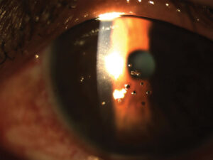
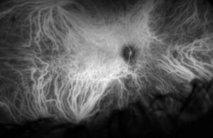
MacTel stages describe how macular telangiectasia type 2 progresses from parafoveal graying to neovascularization.
Gass and Blodi proposed this five-stage grading in 1993. However, OCT now reveals neuroretinal damage before most clinical signs appear, and therefore this guide pairs each stage with its matching imaging findings.
Why MacTel Stages Still Matter
MacTel type 2 is a bilateral neurodegenerative disease of the parafoveal retina that usually presents in middle age with reading difficulty and mild distortion. Because acuity often stays good, patients may arrive with established disease, so staging helps you plan follow-up and explain the outlook.
MacTel Stages at a Glance
Each eye is graded by its most advanced sign, and crystals can appear at any stage, so they do not define a grade.
- Stage 1: near-normal fundus, faint late staining on angiography; OCT may show small inner retinal cavities
- In stage 2, the parafoveal retina looks gray; OCT shows hyporeflective cavities and focal ellipsoid zone loss
- Stage 3: right-angle venules; OCT shows deeper cavitation and outer retinal atrophy
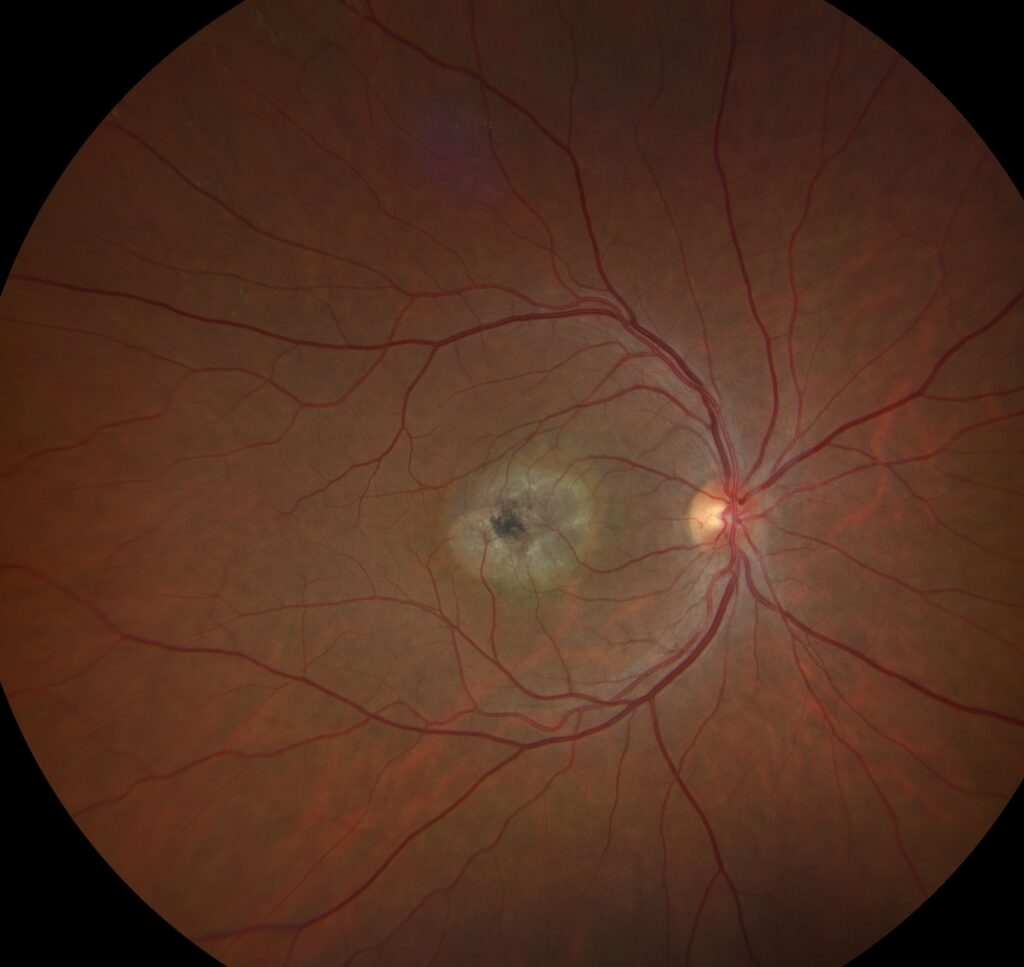
- Stage 4: pigment plaques; OCT shows dense hyperreflective lesions with shadowing
- Finally, stage 5 brings neovascularization; OCT shows hyperreflective material, fluid and a vascular complex
Fundus Explorer Pro
Photograph the retinal findings described here with the phone already in your pocket — 22 D optics and built-in illumination in one handheld unit.
From Choroida — the team behind this siteStage 1: Occult Changes in MacTel Stages
Stage 1 eyes look nearly normal at the slit lamp, and fluorescein angiography may show only mild late staining temporal to the fovea. Meanwhile, macular pigment imaging can already show temporal parafoveal loss, so examine the fellow eye carefully when one eye looks advanced.
Stage 2: Retinal Graying
The first visible sign is a subtle gray loss of parafoveal transparency, which red-free light or a narrow slit beam shows best. On OCT, hyporeflective spaces sit in the inner or outer retina, and unlike diabetic edema, the retina is not thickened.
Stage 3: Right-Angle Venules in MacTel Stages
Stage 3 begins when dilated venules turn sharply and dive into the deep retina, and the vessels look blunted, as if they stop abruptly. OCT angiography shows telangiectatic deep capillaries drawn toward the outer retina. As a result, the ellipsoid zone breaks down over a widening temporal area.
Stage 4: Pigment Plaques
Stage 4 eyes show black intraretinal pigment clumps along the diving venules, and these plaques form when RPE cells migrate inward around the vessels. On OCT, they cast dense shadows over the underlying layers, and their presence signals longstanding disease and a higher neovascular risk.
Stage 5: Neovascularization
Neovascularization defines stage 5, often next to pigment plaques, and patients may notice a sudden drop in vision or new distortion. Next, OCT shows fluid and hyperreflective material around the lesion, and OCT angiography then confirms flow (see macular neovascularization types).
Crystals Across MacTel Stages
Refractile crystals sit in the superficial inner retina and may persist over long periods without change. However, they do not predict progression reliably, and on OCT they appear as tiny hyperreflective dots near the nerve fiber layer.
Where OCT Refines MacTel Stages
The Gass-Blodi grades depend on fundus and angiographic signs, while ellipsoid zone loss on OCT correlates better with visual function. For this reason, the MacTel Project developed multimodal classifications for research and trials, and these newer systems complement the original stages.
Differential Diagnosis of Early MacTel Stages
Diabetic macular edema thickens the retina (see DME OCT patterns), and branch vein occlusion leaves sectoral collaterals and hemorrhage. Lamellar holes also lack graying and right-angle venules, and finally, type 1 MacTel is usually unilateral, with aneurysms and exudate.
Managing Early MacTel Stages
No drug restores vision in nonproliferative disease, and small studies suggest anti-VEGF injections reduce leakage without meaningful visual gain. However, encapsulated cell therapy releasing ciliary neurotrophic factor slowed ellipsoid zone loss in trials, although the implant requires surgery and cannot recover lost photoreceptors.
Treating Stage 5
Anti-VEGF therapy is the standard treatment for neovascular MacTel, and case series suggest many lesions need fewer injections than neovascular AMD. However, atrophy and fibrosis can still limit final acuity.
Counseling Patients
Explain that the disease progresses slowly and rarely causes total blindness. Reading suffers first because damage sits beside the fovea, and you should also check for diabetes, because studies link it with MacTel type 2.
Follow-Up Across MacTel Stages
Many clinicians review early stages yearly with OCT, while stage 4 eyes need closer follow-up because neovascularization often develops near plaques, and you should always image both eyes.
Pitfalls to Avoid
- Do not treat stage 2 cavities as edema
- Never grade by crystals alone
- Do not miss stage 1 in the fellow eye
- Do not assume good acuity means early disease


Document what you see
Two smartphone imaging tools built for everyday clinic use — one for the slit lamp, one for the fundus.
From Choroida — the team behind this siteReferences
- Gass JD, Blodi BA. Idiopathic juxtafoveolar retinal telangiectasis: update of classification and follow-up study. Ophthalmology. 1993;100:1536-1546.
- Yannuzzi LA, Bardal AM, Freund KB, et al. Idiopathic macular telangiectasia. Arch Ophthalmol. 2006;124:450-460.
- Charbel Issa P, Gillies MC, Chew EY, et al. Macular telangiectasia type 2. Prog Retin Eye Res. 2013;34:49-77.
- Chew EY, Clemons TE, Jaffe GJ, et al. Effect of ciliary neurotrophic factor on retinal neurodegeneration in patients with macular telangiectasia type 2: a randomized clinical trial. Ophthalmology. 2019;126:540-549.

Writes and reviews the clinical articles on Educate. Every post is checked against peer-reviewed sources before it goes live.